Chlorine »
PDB 6r6f-6rix »
6rbj »
Chlorine in PDB 6rbj: Crystal Structure of KDM3B in Complex with 5-(1H-Tetrazol-5-Yl) Quinolin-8-Ol
Protein crystallography data
The structure of Crystal Structure of KDM3B in Complex with 5-(1H-Tetrazol-5-Yl) Quinolin-8-Ol, PDB code: 6rbj
was solved by
C.Johansson,
J.A.Newman,
A.Kawamura,
C.J.Schofield,
C.H.Arrowsmith,
C.Bountra,
A.Edwards,
U.C.T.Oppermann,
with X-Ray Crystallography technique. A brief refinement statistics is given in the table below:
| Resolution Low / High (Å) | 86.40 / 2.09 |
| Space group | P 1 21 1 |
| Cell size a, b, c (Å), α, β, γ (°) | 56.929, 93.754, 90.494, 90.00, 107.29, 90.00 |
| R / Rfree (%) | 18.9 / 21 |
Other elements in 6rbj:
The structure of Crystal Structure of KDM3B in Complex with 5-(1H-Tetrazol-5-Yl) Quinolin-8-Ol also contains other interesting chemical elements:
| Manganese | (Mn) | 2 atoms |
Chlorine Binding Sites:
The binding sites of Chlorine atom in the Crystal Structure of KDM3B in Complex with 5-(1H-Tetrazol-5-Yl) Quinolin-8-Ol
(pdb code 6rbj). This binding sites where shown within
5.0 Angstroms radius around Chlorine atom.
In total only one binding site of Chlorine was determined in the Crystal Structure of KDM3B in Complex with 5-(1H-Tetrazol-5-Yl) Quinolin-8-Ol, PDB code: 6rbj:
In total only one binding site of Chlorine was determined in the Crystal Structure of KDM3B in Complex with 5-(1H-Tetrazol-5-Yl) Quinolin-8-Ol, PDB code: 6rbj:
Chlorine binding site 1 out of 1 in 6rbj
Go back to
Chlorine binding site 1 out
of 1 in the Crystal Structure of KDM3B in Complex with 5-(1H-Tetrazol-5-Yl) Quinolin-8-Ol
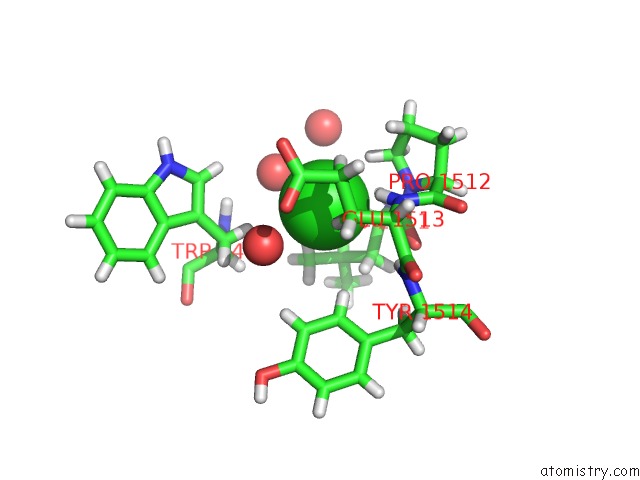
Mono view
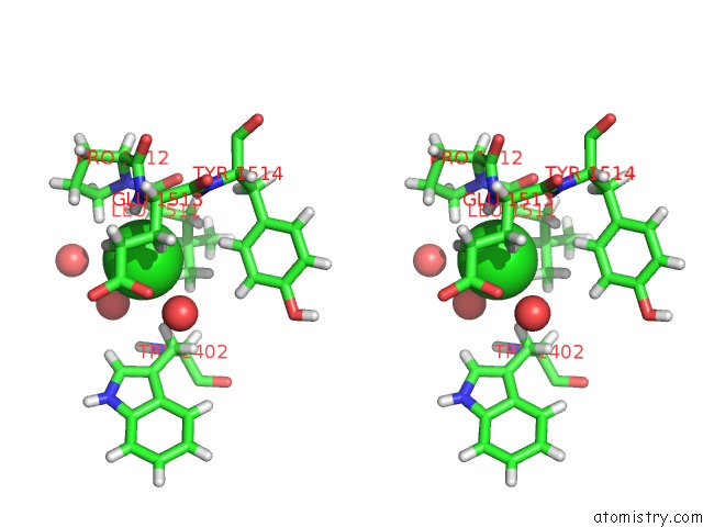
Stereo pair view
Mono view
Stereo pair view
A full contact list of Chlorine with other atoms in the Cl binding
site number 1 of Crystal Structure of KDM3B in Complex with 5-(1H-Tetrazol-5-Yl) Quinolin-8-Ol within 5.0Å range:
|
Reference:
C.Johansson,
J.A.Newman,
A.Kawamura,
C.J.Schofield,
C.H.Arrowsmith,
C.Bountra,
A.Edwards,
U.C.T.Oppermann.
Crystal Structure of KDM3B in Complex with 5-(1H-Tetrazol-5-Yl)Quinolin-8-Ol To Be Published.
Page generated: Sat Jul 12 19:18:23 2025
Last articles
Mg in 4JHDMg in 4JH6
Mg in 4JH8
Mg in 4JH7
Mg in 4JH3
Mg in 4JH5
Mg in 4JF2
Mg in 4JH2
Mg in 4JH1
Mg in 4JEJ