Chlorine »
PDB 6rpn-6s07 »
6rzp »
Chlorine in PDB 6rzp: Multicrystal Structure of Proteinase K at Room Temperature Using A Multilayer Monochromator.
Enzymatic activity of Multicrystal Structure of Proteinase K at Room Temperature Using A Multilayer Monochromator.
All present enzymatic activity of Multicrystal Structure of Proteinase K at Room Temperature Using A Multilayer Monochromator.:
3.4.21.64;
3.4.21.64;
Protein crystallography data
The structure of Multicrystal Structure of Proteinase K at Room Temperature Using A Multilayer Monochromator., PDB code: 6rzp
was solved by
J.Sandy,
E.Sandy,
J.Sanchez-Weatherby,
H.Mikolajek,
with X-Ray Crystallography technique. A brief refinement statistics is given in the table below:
| Resolution Low / High (Å) | 57.13 / 2.20 |
| Space group | P 43 21 2 |
| Cell size a, b, c (Å), α, β, γ (°) | 68.368, 68.368, 103.978, 90.00, 90.00, 90.00 |
| R / Rfree (%) | 10.6 / 16.1 |
Other elements in 6rzp:
The structure of Multicrystal Structure of Proteinase K at Room Temperature Using A Multilayer Monochromator. also contains other interesting chemical elements:
| Calcium | (Ca) | 1 atom |
| Sodium | (Na) | 1 atom |
Chlorine Binding Sites:
The binding sites of Chlorine atom in the Multicrystal Structure of Proteinase K at Room Temperature Using A Multilayer Monochromator.
(pdb code 6rzp). This binding sites where shown within
5.0 Angstroms radius around Chlorine atom.
In total only one binding site of Chlorine was determined in the Multicrystal Structure of Proteinase K at Room Temperature Using A Multilayer Monochromator., PDB code: 6rzp:
In total only one binding site of Chlorine was determined in the Multicrystal Structure of Proteinase K at Room Temperature Using A Multilayer Monochromator., PDB code: 6rzp:
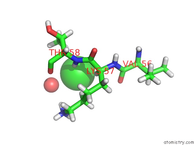
Chlorine binding site 1 out of 1 in 6rzp
Go back to
Chlorine binding site 1 out
of 1 in the Multicrystal Structure of Proteinase K at Room Temperature Using A Multilayer Monochromator.
Mono view
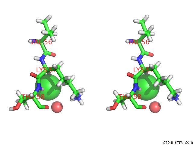
Stereo pair view
Mono view
Stereo pair view
A full contact list of Chlorine with other atoms in the Cl binding
site number 1 of Multicrystal Structure of Proteinase K at Room Temperature Using A Multilayer Monochromator. within 5.0Å range:
|
Reference:
J.Sandy,
E.Sandy,
J.Sanchez-Weatherby,
H.Mikolajek.
Multicrystal Structure of Proteinase K at Room Temperature Using A Multilayer Monochromator. To Be Published.
Page generated: Sat Jul 12 19:34:44 2025
Last articles
Na in 1PH5Na in 1PH4
Na in 1PH3
Na in 1PH6
Na in 1P0Z
Na in 1PA6
Na in 1PH2
Na in 1P9E
Na in 1P59
Na in 1P4Y