Chlorine »
PDB 6sal-6siq »
6set »
Chlorine in PDB 6set: X-Ray Structure of the Gold/Lysozyme Adduct Formed Upon 3 Days Exposure of Protein Crystals to Compound 1
Enzymatic activity of X-Ray Structure of the Gold/Lysozyme Adduct Formed Upon 3 Days Exposure of Protein Crystals to Compound 1
All present enzymatic activity of X-Ray Structure of the Gold/Lysozyme Adduct Formed Upon 3 Days Exposure of Protein Crystals to Compound 1:
3.2.1.17;
3.2.1.17;
Protein crystallography data
The structure of X-Ray Structure of the Gold/Lysozyme Adduct Formed Upon 3 Days Exposure of Protein Crystals to Compound 1, PDB code: 6set
was solved by
G.Ferraro,
A.Giorgio,
A.Merlino,
with X-Ray Crystallography technique. A brief refinement statistics is given in the table below:
| Resolution Low / High (Å) | 30.86 / 1.90 |
| Space group | P 43 21 2 |
| Cell size a, b, c (Å), α, β, γ (°) | 77.448, 77.448, 37.316, 90.00, 90.00, 90.00 |
| R / Rfree (%) | 15.8 / 21.5 |
Other elements in 6set:
The structure of X-Ray Structure of the Gold/Lysozyme Adduct Formed Upon 3 Days Exposure of Protein Crystals to Compound 1 also contains other interesting chemical elements:
| Gold | (Au) | 5 atoms |
| Sodium | (Na) | 1 atom |
Chlorine Binding Sites:
The binding sites of Chlorine atom in the X-Ray Structure of the Gold/Lysozyme Adduct Formed Upon 3 Days Exposure of Protein Crystals to Compound 1
(pdb code 6set). This binding sites where shown within
5.0 Angstroms radius around Chlorine atom.
In total only one binding site of Chlorine was determined in the X-Ray Structure of the Gold/Lysozyme Adduct Formed Upon 3 Days Exposure of Protein Crystals to Compound 1, PDB code: 6set:
In total only one binding site of Chlorine was determined in the X-Ray Structure of the Gold/Lysozyme Adduct Formed Upon 3 Days Exposure of Protein Crystals to Compound 1, PDB code: 6set:
Chlorine binding site 1 out of 1 in 6set
Go back to
Chlorine binding site 1 out
of 1 in the X-Ray Structure of the Gold/Lysozyme Adduct Formed Upon 3 Days Exposure of Protein Crystals to Compound 1
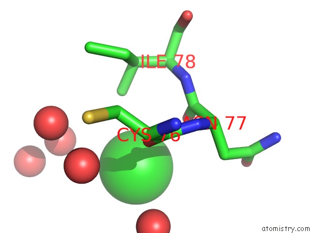
Mono view
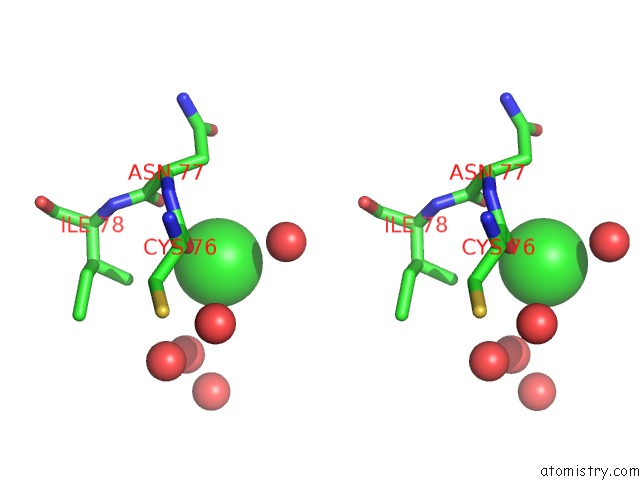
Stereo pair view
Mono view
Stereo pair view
A full contact list of Chlorine with other atoms in the Cl binding
site number 1 of X-Ray Structure of the Gold/Lysozyme Adduct Formed Upon 3 Days Exposure of Protein Crystals to Compound 1 within 5.0Å range:
|
Reference:
G.Ferraro,
A.Giorgio,
A.M.Mansour,
A.Merlino.
Protein-Mediated Disproportionation of Au(I): Insights From the Structures of Adducts of Au(III) Compounds Bearing N,N-Pyridylbenzimidazole Derivatives with Lysozyme. Dalton Trans V. 48 14027 2019.
ISSN: ESSN 1477-9234
PubMed: 31490509
DOI: 10.1039/C9DT02729G
Page generated: Sat Jul 12 19:45:19 2025
ISSN: ESSN 1477-9234
PubMed: 31490509
DOI: 10.1039/C9DT02729G
Last articles
F in 5CI1F in 5CI0
F in 5CGQ
F in 5CDT
F in 5CEP
F in 5CGD
F in 5CGC
F in 5CDS
F in 5CEO
F in 5CDG