Chlorine »
PDB 6sj7-6sr4 »
6slr »
Chlorine in PDB 6slr: Structure of Saposin B in Complex with Atovaquone
Protein crystallography data
The structure of Structure of Saposin B in Complex with Atovaquone, PDB code: 6slr
was solved by
C.Zubieta,
B.Milliken,
R.Doyle,
with X-Ray Crystallography technique. A brief refinement statistics is given in the table below:
| Resolution Low / High (Å) | 38.62 / 2.38 |
| Space group | P 32 2 1 |
| Cell size a, b, c (Å), α, β, γ (°) | 75.707, 75.707, 95.475, 90.00, 90.00, 120.00 |
| R / Rfree (%) | 23.3 / 27.8 |
Chlorine Binding Sites:
The binding sites of Chlorine atom in the Structure of Saposin B in Complex with Atovaquone
(pdb code 6slr). This binding sites where shown within
5.0 Angstroms radius around Chlorine atom.
In total 3 binding sites of Chlorine where determined in the Structure of Saposin B in Complex with Atovaquone, PDB code: 6slr:
Jump to Chlorine binding site number: 1; 2; 3;
In total 3 binding sites of Chlorine where determined in the Structure of Saposin B in Complex with Atovaquone, PDB code: 6slr:
Jump to Chlorine binding site number: 1; 2; 3;
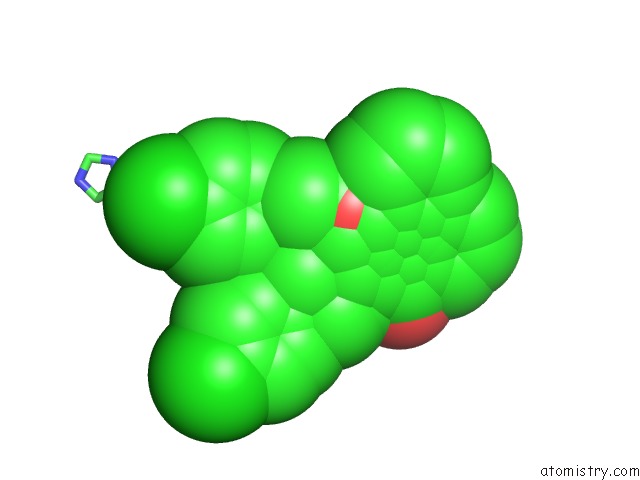
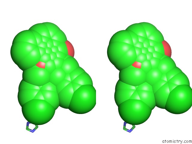
Chlorine binding site 1 out of 3 in 6slr
Go back to
Chlorine binding site 1 out
of 3 in the Structure of Saposin B in Complex with Atovaquone
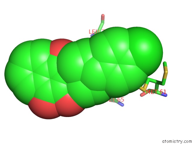
Mono view
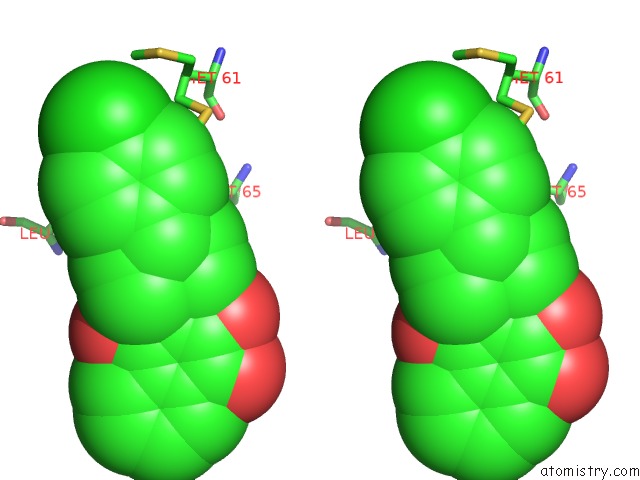
Stereo pair view
Mono view
Stereo pair view
A full contact list of Chlorine with other atoms in the Cl binding
site number 1 of Structure of Saposin B in Complex with Atovaquone within 5.0Å range:
|
Chlorine binding site 2 out of 3 in 6slr
Go back to
Chlorine binding site 2 out
of 3 in the Structure of Saposin B in Complex with Atovaquone
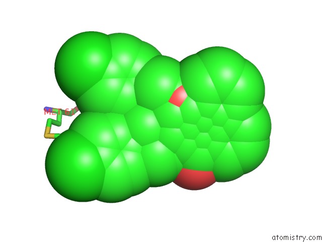
Mono view
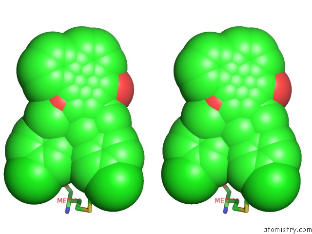
Stereo pair view
Mono view
Stereo pair view
A full contact list of Chlorine with other atoms in the Cl binding
site number 2 of Structure of Saposin B in Complex with Atovaquone within 5.0Å range:
|
Chlorine binding site 3 out of 3 in 6slr
Go back to
Chlorine binding site 3 out
of 3 in the Structure of Saposin B in Complex with Atovaquone
Mono view
Stereo pair view
Mono view
Stereo pair view
A full contact list of Chlorine with other atoms in the Cl binding
site number 3 of Structure of Saposin B in Complex with Atovaquone within 5.0Å range:
|
Reference:
C.Zubieta,
B.Milliken,
R.Doyle.
Structure of Saposin B in Complex with Atovaquone To Be Published.
Page generated: Sat Jul 12 19:49:12 2025
Last articles
Mg in 7UXYMg in 7UY4
Mg in 7UYQ
Mg in 7UXF
Mg in 7UYC
Mg in 7UXW
Mg in 7UXC
Mg in 7UX2
Mg in 7UXA
Mg in 7UUS