Chlorine »
PDB 6tuo-6u5y »
6u30 »
Chlorine in PDB 6u30: The Crystal Structure of 4-Pyridin-3-Ylbenzoate-Bound CYP199A4
Protein crystallography data
The structure of The Crystal Structure of 4-Pyridin-3-Ylbenzoate-Bound CYP199A4, PDB code: 6u30
was solved by
M.N.Podgorski,
J.B.Bruning,
S.G.Bell,
with X-Ray Crystallography technique. A brief refinement statistics is given in the table below:
| Resolution Low / High (Å) | 39.28 / 1.66 |
| Space group | P 1 21 1 |
| Cell size a, b, c (Å), α, β, γ (°) | 44.390, 51.460, 78.820, 90.00, 92.11, 90.00 |
| R / Rfree (%) | 15.1 / 19.3 |
Other elements in 6u30:
The structure of The Crystal Structure of 4-Pyridin-3-Ylbenzoate-Bound CYP199A4 also contains other interesting chemical elements:
| Iron | (Fe) | 1 atom |
Chlorine Binding Sites:
The binding sites of Chlorine atom in the The Crystal Structure of 4-Pyridin-3-Ylbenzoate-Bound CYP199A4
(pdb code 6u30). This binding sites where shown within
5.0 Angstroms radius around Chlorine atom.
In total only one binding site of Chlorine was determined in the The Crystal Structure of 4-Pyridin-3-Ylbenzoate-Bound CYP199A4, PDB code: 6u30:
In total only one binding site of Chlorine was determined in the The Crystal Structure of 4-Pyridin-3-Ylbenzoate-Bound CYP199A4, PDB code: 6u30:
Chlorine binding site 1 out of 1 in 6u30
Go back to
Chlorine binding site 1 out
of 1 in the The Crystal Structure of 4-Pyridin-3-Ylbenzoate-Bound CYP199A4
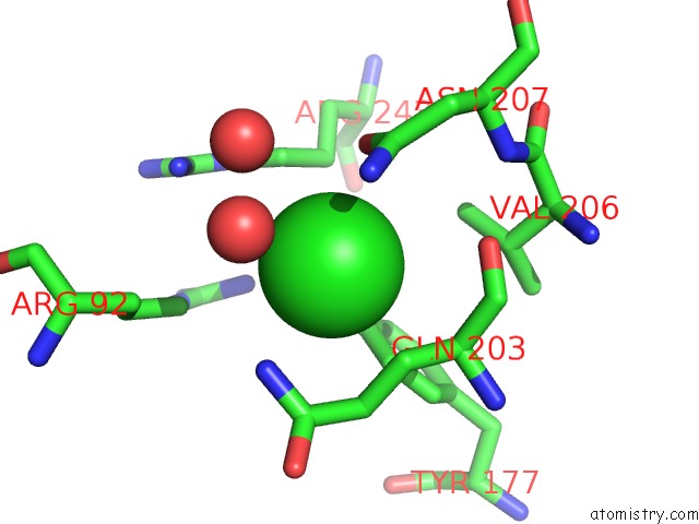
Mono view
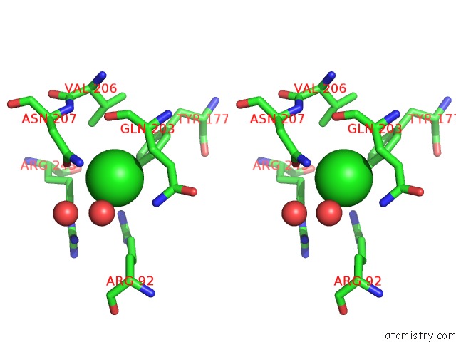
Stereo pair view
Mono view
Stereo pair view
A full contact list of Chlorine with other atoms in the Cl binding
site number 1 of The Crystal Structure of 4-Pyridin-3-Ylbenzoate-Bound CYP199A4 within 5.0Å range:
|
Reference:
M.N.Podgorski,
J.S.Harbort,
T.Coleman,
J.E.Stok,
J.A.Yorke,
L.L.Wong,
J.B.Bruning,
P.V.Bernhardt,
J.J.De Voss,
J.R.Harmer,
S.G.Bell.
Biophysical Techniques to Distinguish Ligand Binding Modes in Cytochrome P450 Monooxygenases Biochemistry 2020.
ISSN: ISSN 0006-2960
Page generated: Sat Jul 12 20:23:05 2025
ISSN: ISSN 0006-2960
Last articles
K in 7QIYK in 7Q3X
K in 7QDN
K in 7QF6
K in 7Q0G
K in 7Q1C
K in 7Q1B
K in 7PXH
K in 7PZJ
K in 7PXG