Chlorine »
PDB 6ury-6v52 »
6usv »
Chlorine in PDB 6usv: Crystal Structure of GLUN1/GLUN2A Ligand-Binding Domain in Complex with Glycine and Sdz 220-040
Protein crystallography data
The structure of Crystal Structure of GLUN1/GLUN2A Ligand-Binding Domain in Complex with Glycine and Sdz 220-040, PDB code: 6usv
was solved by
A.Romero-Hernandez,
N.Tajima,
T.Chou,
H.Furukawa,
with X-Ray Crystallography technique. A brief refinement statistics is given in the table below:
| Resolution Low / High (Å) | 49.50 / 2.30 |
| Space group | P 21 21 21 |
| Cell size a, b, c (Å), α, β, γ (°) | 59.281, 85.131, 121.675, 90.00, 90.00, 90.00 |
| R / Rfree (%) | 19.5 / 25.9 |
Chlorine Binding Sites:
The binding sites of Chlorine atom in the Crystal Structure of GLUN1/GLUN2A Ligand-Binding Domain in Complex with Glycine and Sdz 220-040
(pdb code 6usv). This binding sites where shown within
5.0 Angstroms radius around Chlorine atom.
In total 2 binding sites of Chlorine where determined in the Crystal Structure of GLUN1/GLUN2A Ligand-Binding Domain in Complex with Glycine and Sdz 220-040, PDB code: 6usv:
Jump to Chlorine binding site number: 1; 2;
In total 2 binding sites of Chlorine where determined in the Crystal Structure of GLUN1/GLUN2A Ligand-Binding Domain in Complex with Glycine and Sdz 220-040, PDB code: 6usv:
Jump to Chlorine binding site number: 1; 2;
Chlorine binding site 1 out of 2 in 6usv
Go back to
Chlorine binding site 1 out
of 2 in the Crystal Structure of GLUN1/GLUN2A Ligand-Binding Domain in Complex with Glycine and Sdz 220-040
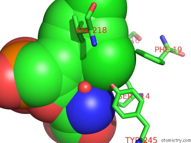
Mono view
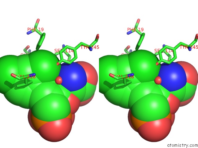
Stereo pair view
Mono view
Stereo pair view
A full contact list of Chlorine with other atoms in the Cl binding
site number 1 of Crystal Structure of GLUN1/GLUN2A Ligand-Binding Domain in Complex with Glycine and Sdz 220-040 within 5.0Å range:
|
Chlorine binding site 2 out of 2 in 6usv
Go back to
Chlorine binding site 2 out
of 2 in the Crystal Structure of GLUN1/GLUN2A Ligand-Binding Domain in Complex with Glycine and Sdz 220-040
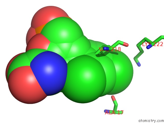
Mono view

Stereo pair view
Mono view
Stereo pair view
A full contact list of Chlorine with other atoms in the Cl binding
site number 2 of Crystal Structure of GLUN1/GLUN2A Ligand-Binding Domain in Complex with Glycine and Sdz 220-040 within 5.0Å range:
|
Reference:
T.H.Chou,
N.Tajima,
A.Romero-Hernandez,
H.Furukawa.
Structural Basis of Functional Transitions in Mammalian Nmda Receptors. Cell V. 182 357 2020.
ISSN: ISSN 1097-4172
PubMed: 32610085
DOI: 10.1016/J.CELL.2020.05.052
Page generated: Sat Jul 12 20:38:07 2025
ISSN: ISSN 1097-4172
PubMed: 32610085
DOI: 10.1016/J.CELL.2020.05.052
Last articles
Na in 3LJQNa in 3LJW
Na in 3LDE
Na in 3LHC
Na in 3LD4
Na in 3LBG
Na in 3KZW
Na in 3L88
Na in 3LA1
Na in 3L9G