Chlorine »
PDB 6vcm-6vkv »
6vcw »
Chlorine in PDB 6vcw: Crystal Structure of Medicago Truncatula S-Adenosylmethionine Synthase 3A (MTMAT3A)
Enzymatic activity of Crystal Structure of Medicago Truncatula S-Adenosylmethionine Synthase 3A (MTMAT3A)
All present enzymatic activity of Crystal Structure of Medicago Truncatula S-Adenosylmethionine Synthase 3A (MTMAT3A):
2.5.1.6;
2.5.1.6;
Protein crystallography data
The structure of Crystal Structure of Medicago Truncatula S-Adenosylmethionine Synthase 3A (MTMAT3A), PDB code: 6vcw
was solved by
B.Sekula,
M.Ruszkowski,
Z.Dauter,
with X-Ray Crystallography technique. A brief refinement statistics is given in the table below:
| Resolution Low / High (Å) | 48.99 / 1.40 |
| Space group | C 1 2 1 |
| Cell size a, b, c (Å), α, β, γ (°) | 111.602, 62.042, 102.284, 90.00, 90.02, 90.00 |
| R / Rfree (%) | 11.5 / 15.9 |
Other elements in 6vcw:
The structure of Crystal Structure of Medicago Truncatula S-Adenosylmethionine Synthase 3A (MTMAT3A) also contains other interesting chemical elements:
| Magnesium | (Mg) | 4 atoms |
Chlorine Binding Sites:
The binding sites of Chlorine atom in the Crystal Structure of Medicago Truncatula S-Adenosylmethionine Synthase 3A (MTMAT3A)
(pdb code 6vcw). This binding sites where shown within
5.0 Angstroms radius around Chlorine atom.
In total 2 binding sites of Chlorine where determined in the Crystal Structure of Medicago Truncatula S-Adenosylmethionine Synthase 3A (MTMAT3A), PDB code: 6vcw:
Jump to Chlorine binding site number: 1; 2;
In total 2 binding sites of Chlorine where determined in the Crystal Structure of Medicago Truncatula S-Adenosylmethionine Synthase 3A (MTMAT3A), PDB code: 6vcw:
Jump to Chlorine binding site number: 1; 2;
Chlorine binding site 1 out of 2 in 6vcw
Go back to
Chlorine binding site 1 out
of 2 in the Crystal Structure of Medicago Truncatula S-Adenosylmethionine Synthase 3A (MTMAT3A)
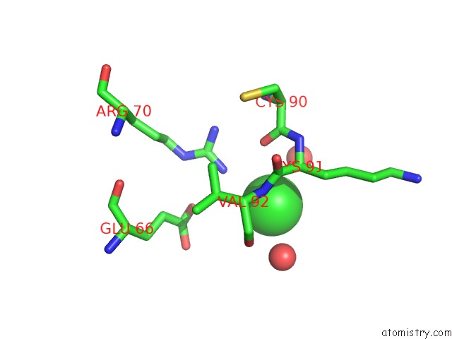
Mono view
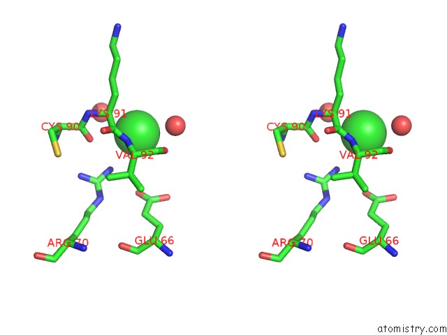
Stereo pair view
Mono view
Stereo pair view
A full contact list of Chlorine with other atoms in the Cl binding
site number 1 of Crystal Structure of Medicago Truncatula S-Adenosylmethionine Synthase 3A (MTMAT3A) within 5.0Å range:
|
Chlorine binding site 2 out of 2 in 6vcw
Go back to
Chlorine binding site 2 out
of 2 in the Crystal Structure of Medicago Truncatula S-Adenosylmethionine Synthase 3A (MTMAT3A)
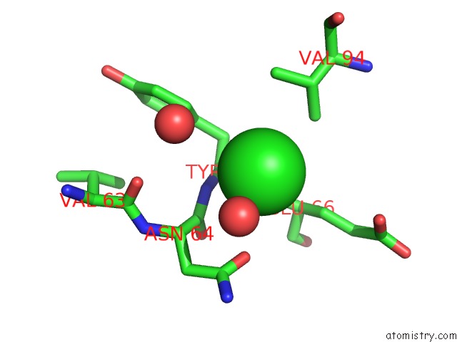
Mono view
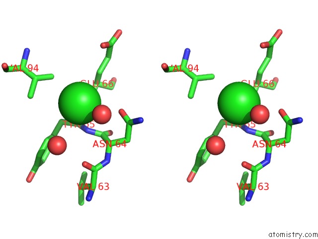
Stereo pair view
Mono view
Stereo pair view
A full contact list of Chlorine with other atoms in the Cl binding
site number 2 of Crystal Structure of Medicago Truncatula S-Adenosylmethionine Synthase 3A (MTMAT3A) within 5.0Å range:
|
Reference:
B.Sekula,
M.Ruszkowski,
Z.Dauter.
S-Adenosylmethionine Synthases in Plants: Structural Characterization of Type I and II Isoenzymes From Arabidopsis Thaliana and Medicago Truncatula. Int.J.Biol.Macromol. 2020.
ISSN: ISSN 0141-8130
PubMed: 32057875
DOI: 10.1016/J.IJBIOMAC.2020.02.100
Page generated: Sat Jul 12 20:50:10 2025
ISSN: ISSN 0141-8130
PubMed: 32057875
DOI: 10.1016/J.IJBIOMAC.2020.02.100
Last articles
K in 8CGUK in 8CGR
K in 8CGJ
K in 8CGI
K in 8CG1
K in 8CFY
K in 8CG2
K in 8CFX
K in 8CG0
K in 8CFW