Chlorine »
PDB 6x6r-6xin »
6xhf »
Chlorine in PDB 6xhf: Structure of Phenylalanyl-5'-O-Adenosine Phosphoramidate
Enzymatic activity of Structure of Phenylalanyl-5'-O-Adenosine Phosphoramidate
All present enzymatic activity of Structure of Phenylalanyl-5'-O-Adenosine Phosphoramidate:
4.6.1.18;
4.6.1.18;
Protein crystallography data
The structure of Structure of Phenylalanyl-5'-O-Adenosine Phosphoramidate, PDB code: 6xhf
was solved by
P.S.Pallan,
M.Egli,
with X-Ray Crystallography technique. A brief refinement statistics is given in the table below:
| Resolution Low / High (Å) | 30.93 / 1.45 |
| Space group | C 1 2 1 |
| Cell size a, b, c (Å), α, β, γ (°) | 100.102, 32.5, 72.945, 90, 90.42, 90 |
| R / Rfree (%) | 19.5 / 23.1 |
Chlorine Binding Sites:
The binding sites of Chlorine atom in the Structure of Phenylalanyl-5'-O-Adenosine Phosphoramidate
(pdb code 6xhf). This binding sites where shown within
5.0 Angstroms radius around Chlorine atom.
In total 2 binding sites of Chlorine where determined in the Structure of Phenylalanyl-5'-O-Adenosine Phosphoramidate, PDB code: 6xhf:
Jump to Chlorine binding site number: 1; 2;
In total 2 binding sites of Chlorine where determined in the Structure of Phenylalanyl-5'-O-Adenosine Phosphoramidate, PDB code: 6xhf:
Jump to Chlorine binding site number: 1; 2;
Chlorine binding site 1 out of 2 in 6xhf
Go back to
Chlorine binding site 1 out
of 2 in the Structure of Phenylalanyl-5'-O-Adenosine Phosphoramidate
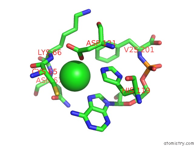
Mono view
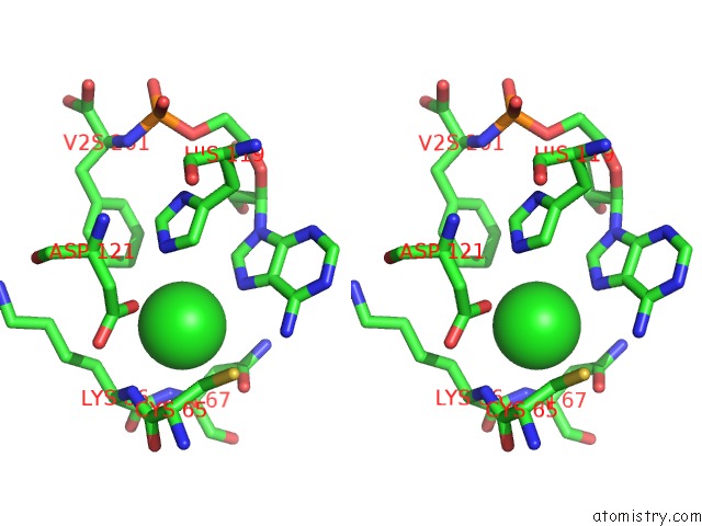
Stereo pair view
Mono view
Stereo pair view
|
|
A full contact list of Chlorine with other atoms in the Cl binding
site number 1 of Structure of Phenylalanyl-5'-O-Adenosine Phosphoramidate within 5.0Å range:
|
Chlorine binding site 2 out of 2 in 6xhf
Go back to
Chlorine binding site 2 out
of 2 in the Structure of Phenylalanyl-5'-O-Adenosine Phosphoramidate
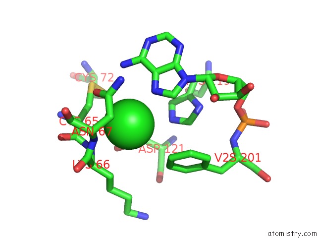
Mono view
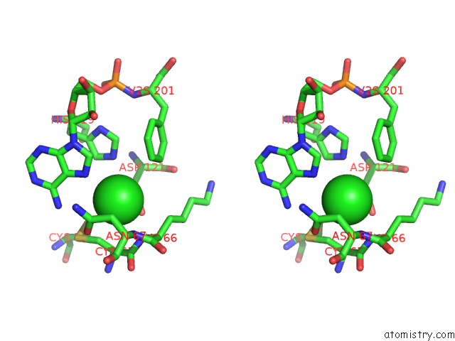
Stereo pair view
Mono view
Stereo pair view
|
|
A full contact list of Chlorine with other atoms in the Cl binding
site number 2 of Structure of Phenylalanyl-5'-O-Adenosine Phosphoramidate within 5.0Å range:
|
Reference:
D.Jovanovic,
P.Tremmel,
P.S.Pallan,
M.Egli,
C.Richert.
The Enzyme-Free Release of Nucleotides From Phosphoramidates Depends Strongly on the Amino Acid. Angew.Chem.Int.Ed.Engl. V. 59 20154 2020.
ISSN: ESSN 1521-3773
PubMed: 32757352
DOI: 10.1002/ANIE.202008665
Page generated: Sat Jul 12 21:39:28 2025
ISSN: ESSN 1521-3773
PubMed: 32757352
DOI: 10.1002/ANIE.202008665
Last articles
Mg in 7A17Mg in 7A0V
Mg in 6ZZX
Mg in 6ZXS
Mg in 7A0Q
Mg in 7A0P
Mg in 7A0C
Mg in 721P
Mg in 6ZXH
Mg in 6ZXG