Chlorine »
PDB 6xz9-6y85 »
6y74 »
Chlorine in PDB 6y74: X-Ray Crystal Structure of Human Carbonic Anhydrase IX Catalytic Domain.
Enzymatic activity of X-Ray Crystal Structure of Human Carbonic Anhydrase IX Catalytic Domain.
All present enzymatic activity of X-Ray Crystal Structure of Human Carbonic Anhydrase IX Catalytic Domain.:
4.2.1.1;
4.2.1.1;
Protein crystallography data
The structure of X-Ray Crystal Structure of Human Carbonic Anhydrase IX Catalytic Domain., PDB code: 6y74
was solved by
S.Z.Fisher,
K.Koruza,
with X-Ray Crystallography technique. A brief refinement statistics is given in the table below:
| Resolution Low / High (Å) | 37.13 / 1.53 |
| Space group | C 1 2 1 |
| Cell size a, b, c (Å), α, β, γ (°) | 113.930, 78.340, 74.320, 90.00, 128.22, 90.00 |
| R / Rfree (%) | 17.4 / 19.8 |
Other elements in 6y74:
The structure of X-Ray Crystal Structure of Human Carbonic Anhydrase IX Catalytic Domain. also contains other interesting chemical elements:
| Zinc | (Zn) | 2 atoms |
Chlorine Binding Sites:
The binding sites of Chlorine atom in the X-Ray Crystal Structure of Human Carbonic Anhydrase IX Catalytic Domain.
(pdb code 6y74). This binding sites where shown within
5.0 Angstroms radius around Chlorine atom.
In total 3 binding sites of Chlorine where determined in the X-Ray Crystal Structure of Human Carbonic Anhydrase IX Catalytic Domain., PDB code: 6y74:
Jump to Chlorine binding site number: 1; 2; 3;
In total 3 binding sites of Chlorine where determined in the X-Ray Crystal Structure of Human Carbonic Anhydrase IX Catalytic Domain., PDB code: 6y74:
Jump to Chlorine binding site number: 1; 2; 3;
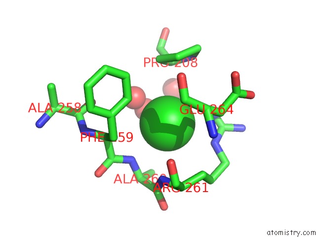
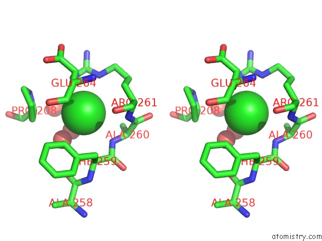
Chlorine binding site 1 out of 3 in 6y74
Go back to
Chlorine binding site 1 out
of 3 in the X-Ray Crystal Structure of Human Carbonic Anhydrase IX Catalytic Domain.
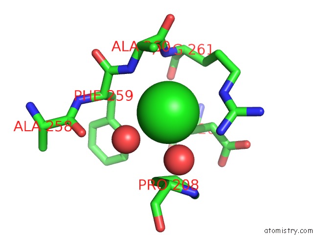
Mono view
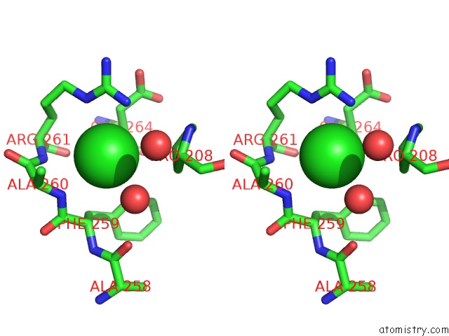
Stereo pair view
Mono view
Stereo pair view
A full contact list of Chlorine with other atoms in the Cl binding
site number 1 of X-Ray Crystal Structure of Human Carbonic Anhydrase IX Catalytic Domain. within 5.0Å range:
|
Chlorine binding site 2 out of 3 in 6y74
Go back to
Chlorine binding site 2 out
of 3 in the X-Ray Crystal Structure of Human Carbonic Anhydrase IX Catalytic Domain.
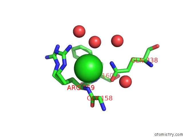
Mono view
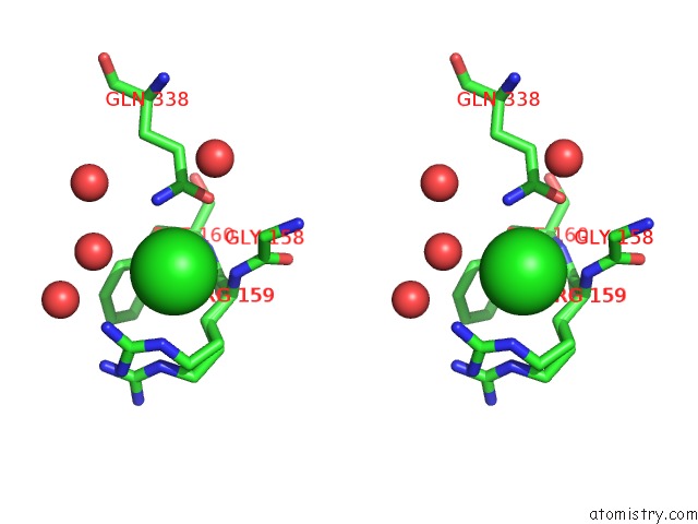
Stereo pair view
Mono view
Stereo pair view
A full contact list of Chlorine with other atoms in the Cl binding
site number 2 of X-Ray Crystal Structure of Human Carbonic Anhydrase IX Catalytic Domain. within 5.0Å range:
|
Chlorine binding site 3 out of 3 in 6y74
Go back to
Chlorine binding site 3 out
of 3 in the X-Ray Crystal Structure of Human Carbonic Anhydrase IX Catalytic Domain.
Mono view
Stereo pair view
Mono view
Stereo pair view
A full contact list of Chlorine with other atoms in the Cl binding
site number 3 of X-Ray Crystal Structure of Human Carbonic Anhydrase IX Catalytic Domain. within 5.0Å range:
|
Reference:
K.Koruza,
A.B.Murray,
B.P.Mahon,
J.B.Hopkins,
W.Knecht,
R.Mckenna,
S.Z.Fisher.
Biophysical Characterization of Cancer-Related Carbonic Anhydrase IX Int J Mol Sci V. 21 5277 2020.
ISSN: ESSN 1422-0067
DOI: 10.3390/IJMS21155277
Page generated: Sat Jul 12 21:54:33 2025
ISSN: ESSN 1422-0067
DOI: 10.3390/IJMS21155277
Last articles
Mg in 1X55Mg in 1X54
Mg in 1X3S
Mg in 1X1T
Mg in 1X1S
Mg in 1X1R
Mg in 1WYW
Mg in 1WVM
Mg in 1X09
Mg in 1X07