Chlorine »
PDB 7aja-7aq0 »
7apm »
Chlorine in PDB 7apm: Trna-Guanine Transglycosylase H319C Mutant Spin-Labeled with Mtsl
Enzymatic activity of Trna-Guanine Transglycosylase H319C Mutant Spin-Labeled with Mtsl
All present enzymatic activity of Trna-Guanine Transglycosylase H319C Mutant Spin-Labeled with Mtsl:
2.4.2.29;
2.4.2.29;
Protein crystallography data
The structure of Trna-Guanine Transglycosylase H319C Mutant Spin-Labeled with Mtsl, PDB code: 7apm
was solved by
D.Nguyen,
A.Heine,
G.Klebe,
with X-Ray Crystallography technique. A brief refinement statistics is given in the table below:
| Resolution Low / High (Å) | 45.68 / 1.66 |
| Space group | C 1 2 1 |
| Cell size a, b, c (Å), α, β, γ (°) | 91.911, 65.064, 70.813, 90.00, 96.29, 90.00 |
| R / Rfree (%) | 13.9 / 17.6 |
Other elements in 7apm:
The structure of Trna-Guanine Transglycosylase H319C Mutant Spin-Labeled with Mtsl also contains other interesting chemical elements:
| Zinc | (Zn) | 1 atom |
Chlorine Binding Sites:
The binding sites of Chlorine atom in the Trna-Guanine Transglycosylase H319C Mutant Spin-Labeled with Mtsl
(pdb code 7apm). This binding sites where shown within
5.0 Angstroms radius around Chlorine atom.
In total only one binding site of Chlorine was determined in the Trna-Guanine Transglycosylase H319C Mutant Spin-Labeled with Mtsl, PDB code: 7apm:
In total only one binding site of Chlorine was determined in the Trna-Guanine Transglycosylase H319C Mutant Spin-Labeled with Mtsl, PDB code: 7apm:
Chlorine binding site 1 out of 1 in 7apm
Go back to
Chlorine binding site 1 out
of 1 in the Trna-Guanine Transglycosylase H319C Mutant Spin-Labeled with Mtsl
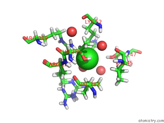
Mono view
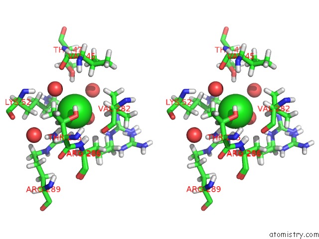
Stereo pair view
Mono view
Stereo pair view
A full contact list of Chlorine with other atoms in the Cl binding
site number 1 of Trna-Guanine Transglycosylase H319C Mutant Spin-Labeled with Mtsl within 5.0Å range:
|
Reference:
D.Abdullin,
D.Nguyen,
C.A.Heubach,
A.Nguyen,
T.Pfaffeneder,
A.Heine,
K.Reuter,
F.Diederich,
O.Schiemann,
G.Klebe.
Unraveling A Ligand-Induced Twist of A Homodimeric Enzyme By Pulsed Electron-Electron Double Resonance To Be Published.
Page generated: Sat Jul 12 22:49:34 2025
Last articles
Fe in 7P63Fe in 7P62
Fe in 7P61
Fe in 7P4Q
Fe in 7P46
Fe in 7P5T
Fe in 7P4M
Fe in 7P4P
Fe in 7P0P
Fe in 7P0R