Chlorine »
PDB 7bar-7bhm »
7bdr »
Chlorine in PDB 7bdr: Structure of Ctx-M-15 E166Q Mutant Crystallised in the Presence of Tazobactam (AAI101)
Enzymatic activity of Structure of Ctx-M-15 E166Q Mutant Crystallised in the Presence of Tazobactam (AAI101)
All present enzymatic activity of Structure of Ctx-M-15 E166Q Mutant Crystallised in the Presence of Tazobactam (AAI101):
3.5.2.6;
3.5.2.6;
Protein crystallography data
The structure of Structure of Ctx-M-15 E166Q Mutant Crystallised in the Presence of Tazobactam (AAI101), PDB code: 7bdr
was solved by
C.L.Tooke,
P.Hinchliffe,
J.Spencer,
with X-Ray Crystallography technique. A brief refinement statistics is given in the table below:
| Resolution Low / High (Å) | 42.56 / 0.91 |
| Space group | P 21 21 21 |
| Cell size a, b, c (Å), α, β, γ (°) | 44.678, 45.671, 117.33, 90, 90, 90 |
| R / Rfree (%) | 11.4 / 12.6 |
Other elements in 7bdr:
The structure of Structure of Ctx-M-15 E166Q Mutant Crystallised in the Presence of Tazobactam (AAI101) also contains other interesting chemical elements:
| Sodium | (Na) | 1 atom |
Chlorine Binding Sites:
The binding sites of Chlorine atom in the Structure of Ctx-M-15 E166Q Mutant Crystallised in the Presence of Tazobactam (AAI101)
(pdb code 7bdr). This binding sites where shown within
5.0 Angstroms radius around Chlorine atom.
In total only one binding site of Chlorine was determined in the Structure of Ctx-M-15 E166Q Mutant Crystallised in the Presence of Tazobactam (AAI101), PDB code: 7bdr:
In total only one binding site of Chlorine was determined in the Structure of Ctx-M-15 E166Q Mutant Crystallised in the Presence of Tazobactam (AAI101), PDB code: 7bdr:
Chlorine binding site 1 out of 1 in 7bdr
Go back to
Chlorine binding site 1 out
of 1 in the Structure of Ctx-M-15 E166Q Mutant Crystallised in the Presence of Tazobactam (AAI101)
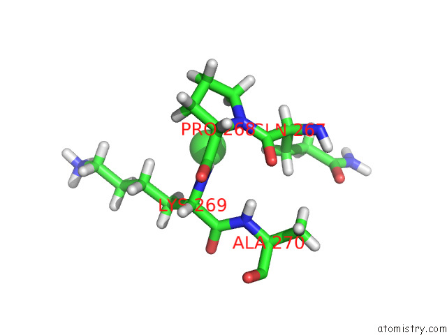
Mono view
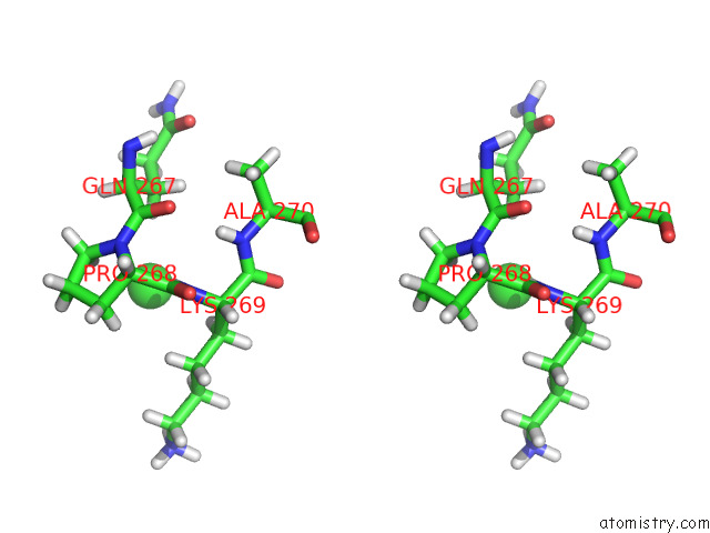
Stereo pair view
Mono view
Stereo pair view
A full contact list of Chlorine with other atoms in the Cl binding
site number 1 of Structure of Ctx-M-15 E166Q Mutant Crystallised in the Presence of Tazobactam (AAI101) within 5.0Å range:
|
Reference:
P.Hinchliffe,
C.L.Tooke,
C.R.Bethel,
B.Wang,
C.Arthur,
K.J.Heesom,
S.Shapiro,
D.M.Schlatzer,
K.M.Papp-Wallace,
R.A.Bonomo,
J.Spencer.
Penicillanic Acid Sulfones Inactivate the Extended-Spectrum Beta-Lactamase Ctx-M-15 Through Formation of A Serine-Lysine Cross-Link: An Alternative Mechanism of Beta-Lactamase Inhibition. Mbio V. 13 79321 2022.
ISSN: ESSN 2150-7511
PubMed: 35612361
DOI: 10.1128/MBIO.01793-21
Page generated: Sat Jul 12 23:15:17 2025
ISSN: ESSN 2150-7511
PubMed: 35612361
DOI: 10.1128/MBIO.01793-21
Last articles
Fe in 2YXOFe in 2YRS
Fe in 2YXC
Fe in 2YNM
Fe in 2YVJ
Fe in 2YP1
Fe in 2YU2
Fe in 2YU1
Fe in 2YQB
Fe in 2YOO