Chlorine »
PDB 7c3a-7c7b »
7c5h »
Chlorine in PDB 7c5h: Crystal Structure of Glyceraldehyde-3-Phosphate DEHYDROGENASE1 From Escherichia Coli at 2.09 Angstrom Resolution
Protein crystallography data
The structure of Crystal Structure of Glyceraldehyde-3-Phosphate DEHYDROGENASE1 From Escherichia Coli at 2.09 Angstrom Resolution, PDB code: 7c5h
was solved by
L.Zhang,
M.R.Liu,
L.Y.Bao,
Y.C.Yao,
I.K.Bostrom,
Y.D.Wang,
A.Q.Chen,
J.X.Li,
S.H.Gu,
C.N.Ji,
with X-Ray Crystallography technique. A brief refinement statistics is given in the table below:
| Resolution Low / High (Å) | 40.00 / 2.09 |
| Space group | P 41 21 2 |
| Cell size a, b, c (Å), α, β, γ (°) | 89.778, 89.778, 340.953, 90, 90, 90 |
| R / Rfree (%) | 15.1 / 20.3 |
Chlorine Binding Sites:
The binding sites of Chlorine atom in the Crystal Structure of Glyceraldehyde-3-Phosphate DEHYDROGENASE1 From Escherichia Coli at 2.09 Angstrom Resolution
(pdb code 7c5h). This binding sites where shown within
5.0 Angstroms radius around Chlorine atom.
In total 7 binding sites of Chlorine where determined in the Crystal Structure of Glyceraldehyde-3-Phosphate DEHYDROGENASE1 From Escherichia Coli at 2.09 Angstrom Resolution, PDB code: 7c5h:
Jump to Chlorine binding site number: 1; 2; 3; 4; 5; 6; 7;
In total 7 binding sites of Chlorine where determined in the Crystal Structure of Glyceraldehyde-3-Phosphate DEHYDROGENASE1 From Escherichia Coli at 2.09 Angstrom Resolution, PDB code: 7c5h:
Jump to Chlorine binding site number: 1; 2; 3; 4; 5; 6; 7;
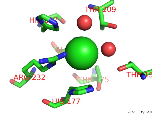
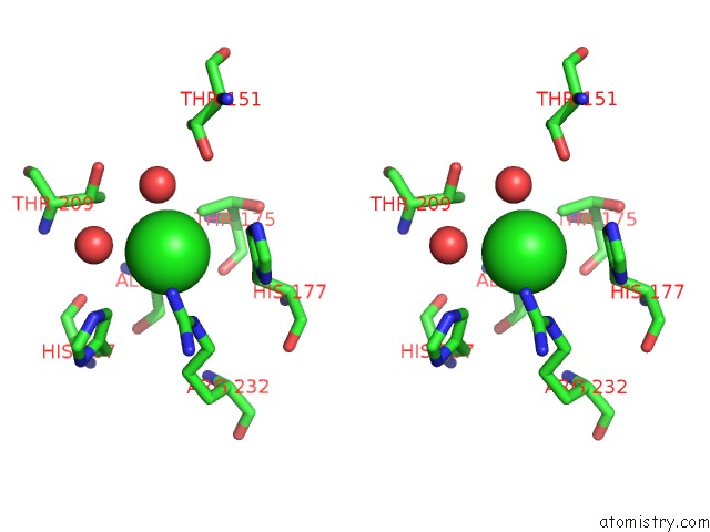
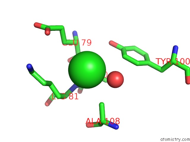
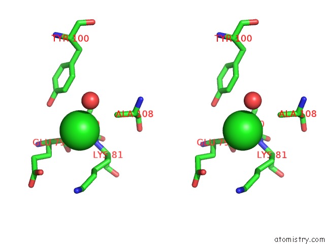
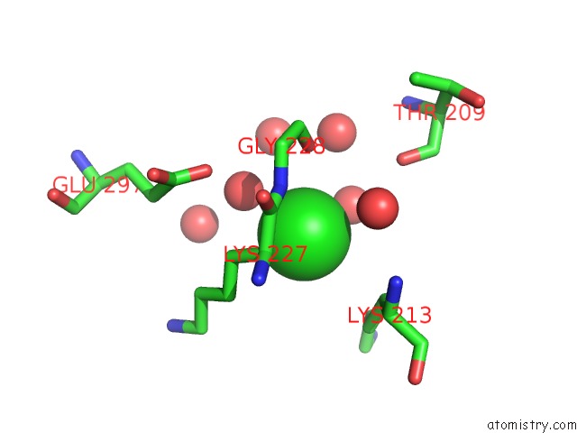
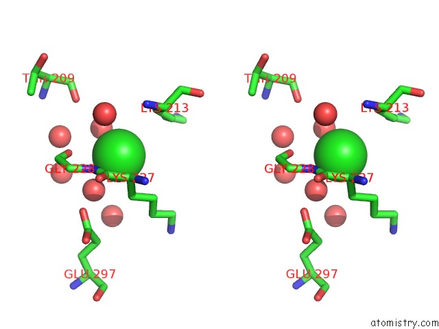
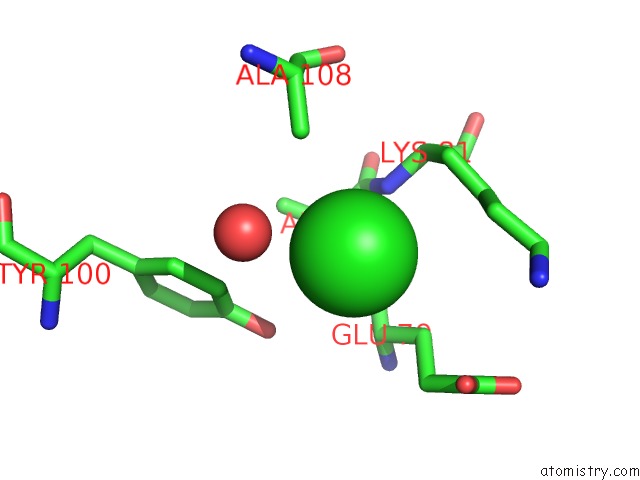
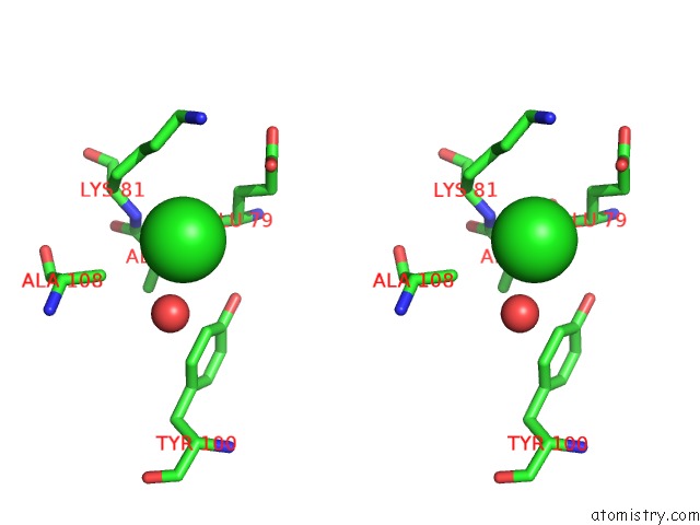
Chlorine binding site 1 out of 7 in 7c5h
Go back to
Chlorine binding site 1 out
of 7 in the Crystal Structure of Glyceraldehyde-3-Phosphate DEHYDROGENASE1 From Escherichia Coli at 2.09 Angstrom Resolution
Mono view
Stereo pair view
Mono view
Stereo pair view
A full contact list of Chlorine with other atoms in the Cl binding
site number 1 of Crystal Structure of Glyceraldehyde-3-Phosphate DEHYDROGENASE1 From Escherichia Coli at 2.09 Angstrom Resolution within 5.0Å range:
|
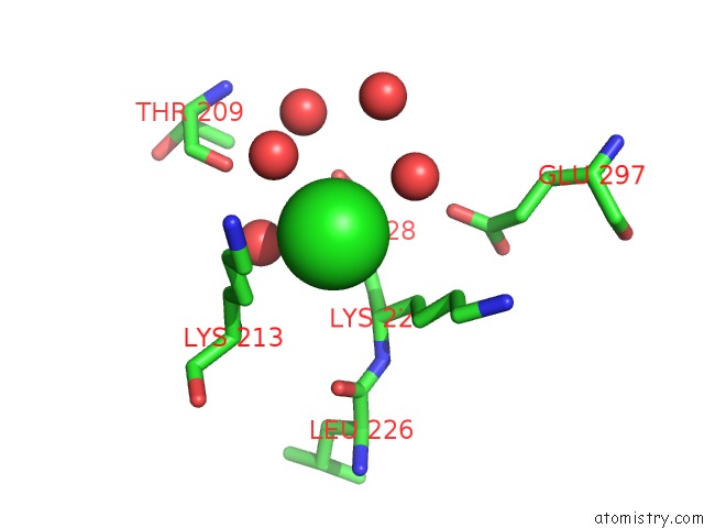
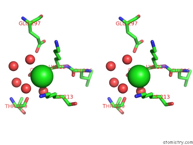
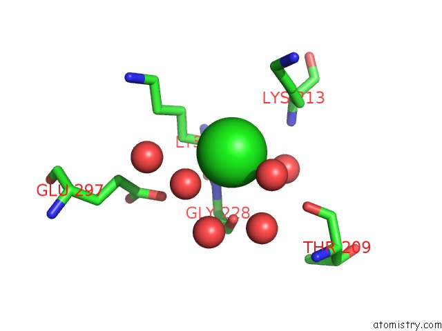
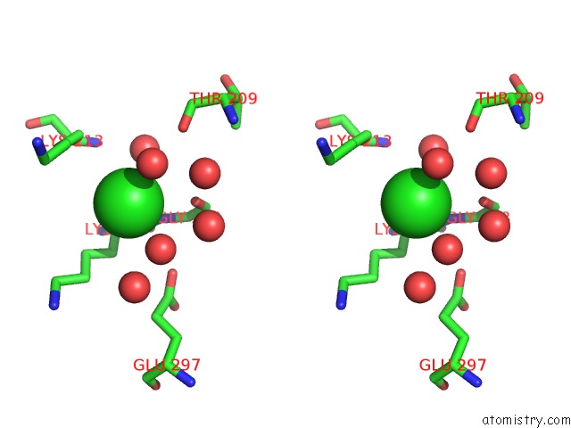
Chlorine binding site 2 out of 7 in 7c5h
Go back to
Chlorine binding site 2 out
of 7 in the Crystal Structure of Glyceraldehyde-3-Phosphate DEHYDROGENASE1 From Escherichia Coli at 2.09 Angstrom Resolution
Mono view
Stereo pair view
Mono view
Stereo pair view
A full contact list of Chlorine with other atoms in the Cl binding
site number 2 of Crystal Structure of Glyceraldehyde-3-Phosphate DEHYDROGENASE1 From Escherichia Coli at 2.09 Angstrom Resolution within 5.0Å range:
|
Chlorine binding site 3 out of 7 in 7c5h
Go back to
Chlorine binding site 3 out
of 7 in the Crystal Structure of Glyceraldehyde-3-Phosphate DEHYDROGENASE1 From Escherichia Coli at 2.09 Angstrom Resolution
Mono view
Stereo pair view
Mono view
Stereo pair view
A full contact list of Chlorine with other atoms in the Cl binding
site number 3 of Crystal Structure of Glyceraldehyde-3-Phosphate DEHYDROGENASE1 From Escherichia Coli at 2.09 Angstrom Resolution within 5.0Å range:
|
Chlorine binding site 4 out of 7 in 7c5h
Go back to
Chlorine binding site 4 out
of 7 in the Crystal Structure of Glyceraldehyde-3-Phosphate DEHYDROGENASE1 From Escherichia Coli at 2.09 Angstrom Resolution
Mono view
Stereo pair view
Mono view
Stereo pair view
A full contact list of Chlorine with other atoms in the Cl binding
site number 4 of Crystal Structure of Glyceraldehyde-3-Phosphate DEHYDROGENASE1 From Escherichia Coli at 2.09 Angstrom Resolution within 5.0Å range:
|
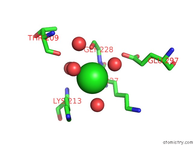
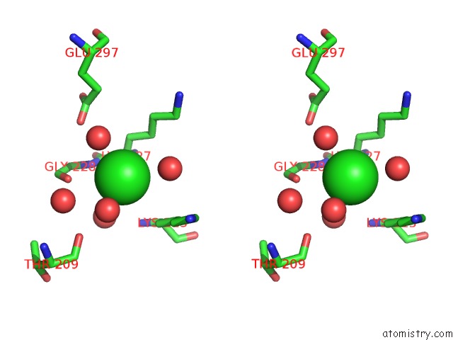
Chlorine binding site 5 out of 7 in 7c5h
Go back to
Chlorine binding site 5 out
of 7 in the Crystal Structure of Glyceraldehyde-3-Phosphate DEHYDROGENASE1 From Escherichia Coli at 2.09 Angstrom Resolution
Mono view
Stereo pair view
Mono view
Stereo pair view
A full contact list of Chlorine with other atoms in the Cl binding
site number 5 of Crystal Structure of Glyceraldehyde-3-Phosphate DEHYDROGENASE1 From Escherichia Coli at 2.09 Angstrom Resolution within 5.0Å range:
|
Chlorine binding site 6 out of 7 in 7c5h
Go back to
Chlorine binding site 6 out
of 7 in the Crystal Structure of Glyceraldehyde-3-Phosphate DEHYDROGENASE1 From Escherichia Coli at 2.09 Angstrom Resolution
Mono view
Stereo pair view
Mono view
Stereo pair view
A full contact list of Chlorine with other atoms in the Cl binding
site number 6 of Crystal Structure of Glyceraldehyde-3-Phosphate DEHYDROGENASE1 From Escherichia Coli at 2.09 Angstrom Resolution within 5.0Å range:
|
Chlorine binding site 7 out of 7 in 7c5h
Go back to
Chlorine binding site 7 out
of 7 in the Crystal Structure of Glyceraldehyde-3-Phosphate DEHYDROGENASE1 From Escherichia Coli at 2.09 Angstrom Resolution
Mono view
Stereo pair view
Mono view
Stereo pair view
A full contact list of Chlorine with other atoms in the Cl binding
site number 7 of Crystal Structure of Glyceraldehyde-3-Phosphate DEHYDROGENASE1 From Escherichia Coli at 2.09 Angstrom Resolution within 5.0Å range:
|
Reference:
L.Zhang,
M.R.Liu,
L.Y.Bao,
Y.C.Yao,
I.K.Bostrom,
Y.D.Wang,
A.Q.Chen,
J.X.Li,
S.H.Gu,
C.N.Ji.
Crystal Structure of TYPE1 Glyceraldehyde-3-Phosphate Dehydrogenase From Escherichia Coli Provides New Insight Into Bpg Generation and Catalytic Mechanism To Be Published.
Page generated: Sat Jul 12 23:31:51 2025
Last articles
K in 1YJ9K in 1YJ3
K in 1YIJ
K in 1YI2
K in 1YIT
K in 1YHQ
K in 1Y8P
K in 1Y8O
K in 1Y3S
K in 1Y39