Chlorine »
PDB 7e4q-7epg »
7e5u »
Chlorine in PDB 7e5u: Crystal Structure of PHM7
Protein crystallography data
The structure of Crystal Structure of PHM7, PDB code: 7e5u
was solved by
K.Fujiyama,
N.Kato,
K.Kinugasa,
T.Hino,
S.Takahashi,
S.Nagano,
with X-Ray Crystallography technique. A brief refinement statistics is given in the table below:
| Resolution Low / High (Å) | 43.87 / 1.62 |
| Space group | C 1 2 1 |
| Cell size a, b, c (Å), α, β, γ (°) | 91.206, 150.481, 99.333, 90, 97.35, 90 |
| R / Rfree (%) | 18.9 / 22.2 |
Chlorine Binding Sites:
The binding sites of Chlorine atom in the Crystal Structure of PHM7
(pdb code 7e5u). This binding sites where shown within
5.0 Angstroms radius around Chlorine atom.
In total 2 binding sites of Chlorine where determined in the Crystal Structure of PHM7, PDB code: 7e5u:
Jump to Chlorine binding site number: 1; 2;
In total 2 binding sites of Chlorine where determined in the Crystal Structure of PHM7, PDB code: 7e5u:
Jump to Chlorine binding site number: 1; 2;
Chlorine binding site 1 out of 2 in 7e5u
Go back to
Chlorine binding site 1 out
of 2 in the Crystal Structure of PHM7
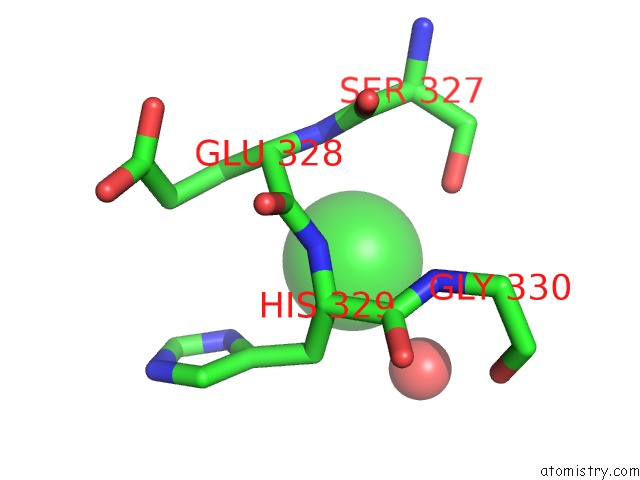
Mono view
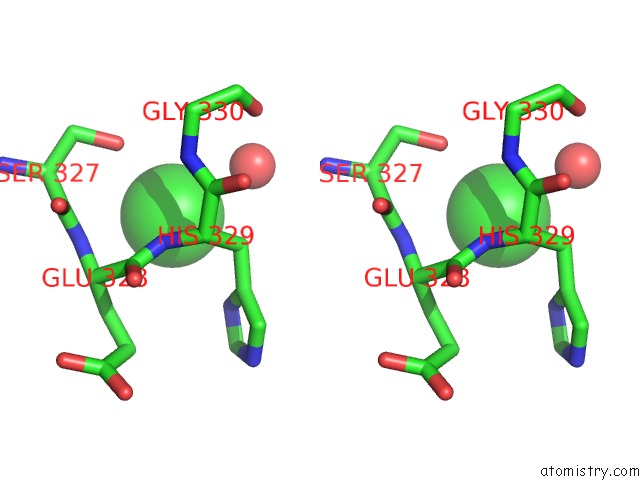
Stereo pair view
Mono view
Stereo pair view
A full contact list of Chlorine with other atoms in the Cl binding
site number 1 of Crystal Structure of PHM7 within 5.0Å range:
|
Chlorine binding site 2 out of 2 in 7e5u
Go back to
Chlorine binding site 2 out
of 2 in the Crystal Structure of PHM7
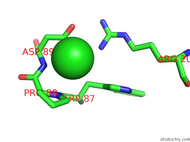
Mono view
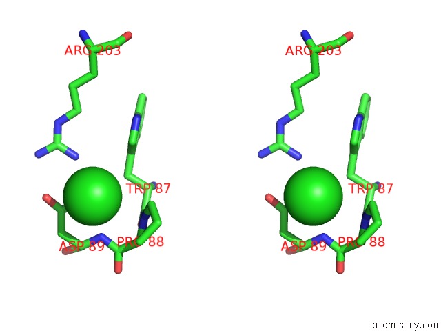
Stereo pair view
Mono view
Stereo pair view
A full contact list of Chlorine with other atoms in the Cl binding
site number 2 of Crystal Structure of PHM7 within 5.0Å range:
|
Reference:
K.Fujiyama,
N.Kato,
S.Re,
K.Kinugasa,
K.Watanabe,
R.Takita,
T.Nogawa,
T.Hino,
H.Osada,
Y.Sugita,
S.Takahashi,
S.Nagano.
Molecular Basis For Two Stereoselective Diels-Alderases That Produce Decalin Skeletons. Angew.Chem.Int.Ed.Engl. 2021.
ISSN: ESSN 1521-3773
PubMed: 34121297
DOI: 10.1002/ANIE.202106186
Page generated: Sun Jul 13 00:20:28 2025
ISSN: ESSN 1521-3773
PubMed: 34121297
DOI: 10.1002/ANIE.202106186
Last articles
Mn in 9LJUMn in 9LJW
Mn in 9LJS
Mn in 9LJR
Mn in 9LJT
Mn in 9LJV
Mg in 9UA2
Mg in 9R96
Mg in 9VM1
Mg in 9P01