Chlorine »
PDB 7epi-7f6k »
7epj »
Chlorine in PDB 7epj: Crystal Structure of E.Coli Ccdb Mutant V46L
Protein crystallography data
The structure of Crystal Structure of E.Coli Ccdb Mutant V46L, PDB code: 7epj
was solved by
K.Manjunath,
P.Goyal,
R.Varadarajan,
with X-Ray Crystallography technique. A brief refinement statistics is given in the table below:
| Resolution Low / High (Å) | 33.98 / 1.35 |
| Space group | C 1 2 1 |
| Cell size a, b, c (Å), α, β, γ (°) | 75.1, 36.76, 35.91, 90, 115.19, 90 |
| R / Rfree (%) | 16.2 / 18.4 |
Chlorine Binding Sites:
The binding sites of Chlorine atom in the Crystal Structure of E.Coli Ccdb Mutant V46L
(pdb code 7epj). This binding sites where shown within
5.0 Angstroms radius around Chlorine atom.
In total 2 binding sites of Chlorine where determined in the Crystal Structure of E.Coli Ccdb Mutant V46L, PDB code: 7epj:
Jump to Chlorine binding site number: 1; 2;
In total 2 binding sites of Chlorine where determined in the Crystal Structure of E.Coli Ccdb Mutant V46L, PDB code: 7epj:
Jump to Chlorine binding site number: 1; 2;
Chlorine binding site 1 out of 2 in 7epj
Go back to
Chlorine binding site 1 out
of 2 in the Crystal Structure of E.Coli Ccdb Mutant V46L
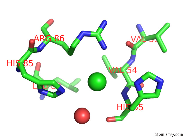
Mono view
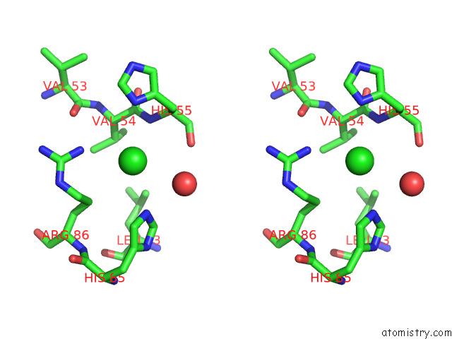
Stereo pair view
Mono view
Stereo pair view
A full contact list of Chlorine with other atoms in the Cl binding
site number 1 of Crystal Structure of E.Coli Ccdb Mutant V46L within 5.0Å range:
|
Chlorine binding site 2 out of 2 in 7epj
Go back to
Chlorine binding site 2 out
of 2 in the Crystal Structure of E.Coli Ccdb Mutant V46L
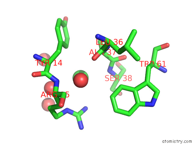
Mono view
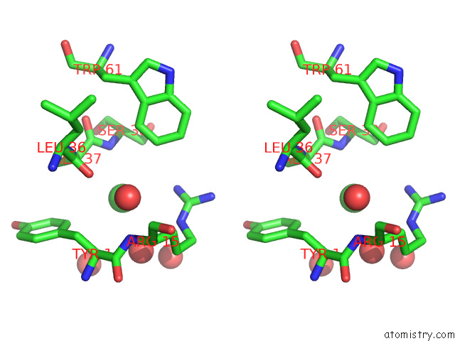
Stereo pair view
Mono view
Stereo pair view
A full contact list of Chlorine with other atoms in the Cl binding
site number 2 of Crystal Structure of E.Coli Ccdb Mutant V46L within 5.0Å range:
|
Reference:
G.Chattopadhyay,
J.Bhowmick,
K.Manjunath,
S.Ahmed,
P.Goyal,
R.Varadarajan.
Mechanistic Insights Into Global Suppressors of Protein Folding Defects. Plos Genet. V. 18 10334 2022.
ISSN: ESSN 1553-7404
PubMed: 36037221
DOI: 10.1371/JOURNAL.PGEN.1010334
Page generated: Sun Jul 13 00:26:56 2025
ISSN: ESSN 1553-7404
PubMed: 36037221
DOI: 10.1371/JOURNAL.PGEN.1010334
Last articles
Mn in 5TYWMn in 5TYV
Mn in 5TIS
Mn in 5TYU
Mn in 5TWQ
Mn in 5TIR
Mn in 5TG3
Mn in 5TSN
Mn in 5TB9
Mn in 5TB8