Chlorine »
PDB 7epi-7f6k »
7er3 »
Chlorine in PDB 7er3: Crystal Structure of Beta-Lactoglobulin Complexed with Chloroquine
Protein crystallography data
The structure of Crystal Structure of Beta-Lactoglobulin Complexed with Chloroquine, PDB code: 7er3
was solved by
Q.Yao,
J.Ma,
Y.Xing,
J.Zang,
with X-Ray Crystallography technique. A brief refinement statistics is given in the table below:
| Resolution Low / High (Å) | 29.60 / 2.60 |
| Space group | C 1 2 1 |
| Cell size a, b, c (Å), α, β, γ (°) | 150.118, 66.953, 78.044, 90, 111.74, 90 |
| R / Rfree (%) | 24.7 / 32.4 |
Chlorine Binding Sites:
The binding sites of Chlorine atom in the Crystal Structure of Beta-Lactoglobulin Complexed with Chloroquine
(pdb code 7er3). This binding sites where shown within
5.0 Angstroms radius around Chlorine atom.
In total 4 binding sites of Chlorine where determined in the Crystal Structure of Beta-Lactoglobulin Complexed with Chloroquine, PDB code: 7er3:
Jump to Chlorine binding site number: 1; 2; 3; 4;
In total 4 binding sites of Chlorine where determined in the Crystal Structure of Beta-Lactoglobulin Complexed with Chloroquine, PDB code: 7er3:
Jump to Chlorine binding site number: 1; 2; 3; 4;
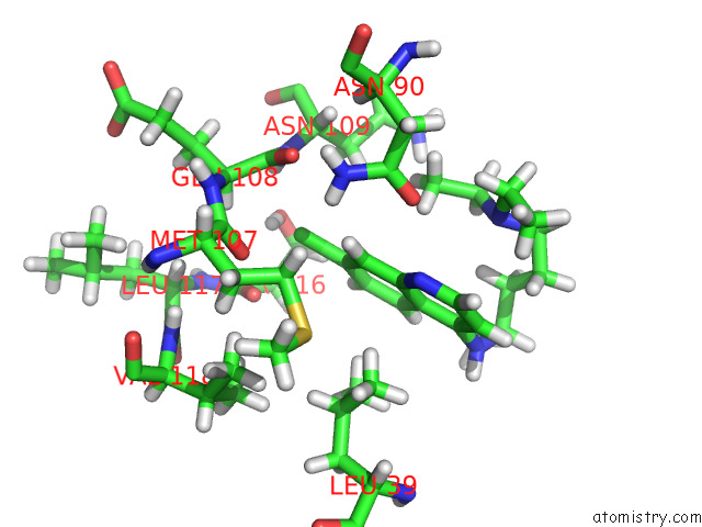
Chlorine binding site 1 out of 4 in 7er3
Go back to
Chlorine binding site 1 out
of 4 in the Crystal Structure of Beta-Lactoglobulin Complexed with Chloroquine
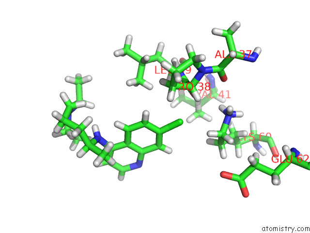
Mono view
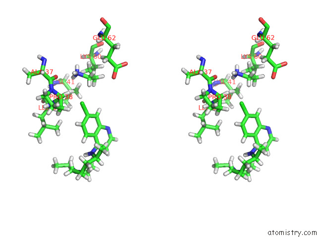
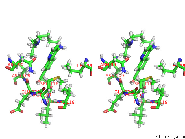
Stereo pair view
Mono view
Stereo pair view
A full contact list of Chlorine with other atoms in the Cl binding
site number 1 of Crystal Structure of Beta-Lactoglobulin Complexed with Chloroquine within 5.0Å range:
|
Chlorine binding site 2 out of 4 in 7er3
Go back to
Chlorine binding site 2 out
of 4 in the Crystal Structure of Beta-Lactoglobulin Complexed with Chloroquine
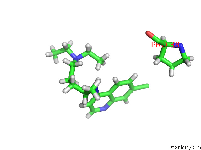
Mono view
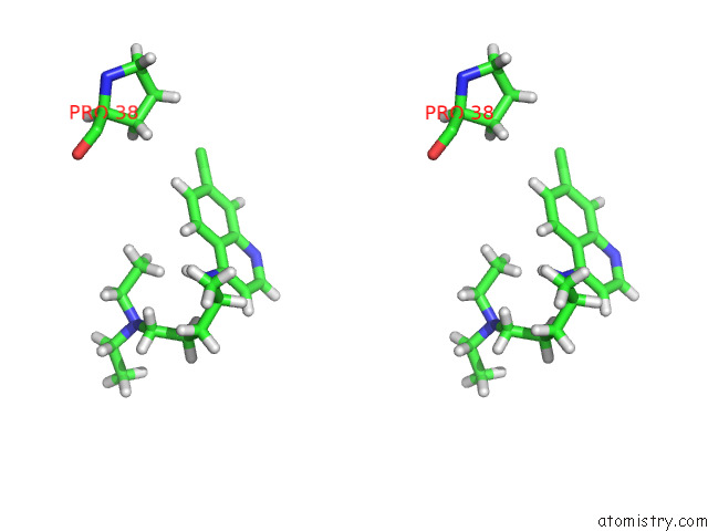
Stereo pair view
Mono view
Stereo pair view
A full contact list of Chlorine with other atoms in the Cl binding
site number 2 of Crystal Structure of Beta-Lactoglobulin Complexed with Chloroquine within 5.0Å range:
|
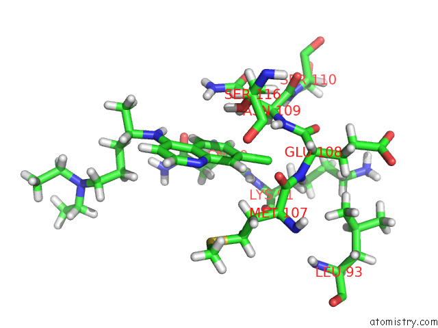
Chlorine binding site 3 out of 4 in 7er3
Go back to
Chlorine binding site 3 out
of 4 in the Crystal Structure of Beta-Lactoglobulin Complexed with Chloroquine
Mono view
Stereo pair view
Mono view
Stereo pair view
A full contact list of Chlorine with other atoms in the Cl binding
site number 3 of Crystal Structure of Beta-Lactoglobulin Complexed with Chloroquine within 5.0Å range:
|
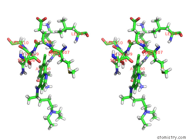
Chlorine binding site 4 out of 4 in 7er3
Go back to
Chlorine binding site 4 out
of 4 in the Crystal Structure of Beta-Lactoglobulin Complexed with Chloroquine
Mono view
Stereo pair view
Mono view
Stereo pair view
A full contact list of Chlorine with other atoms in the Cl binding
site number 4 of Crystal Structure of Beta-Lactoglobulin Complexed with Chloroquine within 5.0Å range:
|
Reference:
Q.Yao,
J.Ma,
Y.Xing,
J.Zang.
Crystal Structure of Beta-Lactoglobulin Complexed with Chloroquine To Be Published.
Page generated: Sun Jul 13 00:27:26 2025
Last articles
Na in 6OL1Na in 6ONT
Na in 6OOG
Na in 6ON9
Na in 6OKZ
Na in 6OL0
Na in 6OME
Na in 6OL8
Na in 6OKF
Na in 6OFD