Chlorine »
PDB 7f6q-7fgy »
7far »
Chlorine in PDB 7far: Crystal Structure of PDE5A in Complex with Inhibitor L12
Enzymatic activity of Crystal Structure of PDE5A in Complex with Inhibitor L12
All present enzymatic activity of Crystal Structure of PDE5A in Complex with Inhibitor L12:
3.1.4.35;
3.1.4.35;
Protein crystallography data
The structure of Crystal Structure of PDE5A in Complex with Inhibitor L12, PDB code: 7far
was solved by
D.Wu,
Y.Y.Huang,
H.B.Luo,
with X-Ray Crystallography technique. A brief refinement statistics is given in the table below:
| Resolution Low / High (Å) | 23.80 / 2.40 |
| Space group | P 31 2 1 |
| Cell size a, b, c (Å), α, β, γ (°) | 73.917, 73.917, 132.251, 90, 90, 120 |
| R / Rfree (%) | 21.3 / 25 |
Other elements in 7far:
The structure of Crystal Structure of PDE5A in Complex with Inhibitor L12 also contains other interesting chemical elements:
| Fluorine | (F) | 2 atoms |
| Magnesium | (Mg) | 1 atom |
| Zinc | (Zn) | 1 atom |
Chlorine Binding Sites:
The binding sites of Chlorine atom in the Crystal Structure of PDE5A in Complex with Inhibitor L12
(pdb code 7far). This binding sites where shown within
5.0 Angstroms radius around Chlorine atom.
In total only one binding site of Chlorine was determined in the Crystal Structure of PDE5A in Complex with Inhibitor L12, PDB code: 7far:
In total only one binding site of Chlorine was determined in the Crystal Structure of PDE5A in Complex with Inhibitor L12, PDB code: 7far:
Chlorine binding site 1 out of 1 in 7far
Go back to
Chlorine binding site 1 out
of 1 in the Crystal Structure of PDE5A in Complex with Inhibitor L12
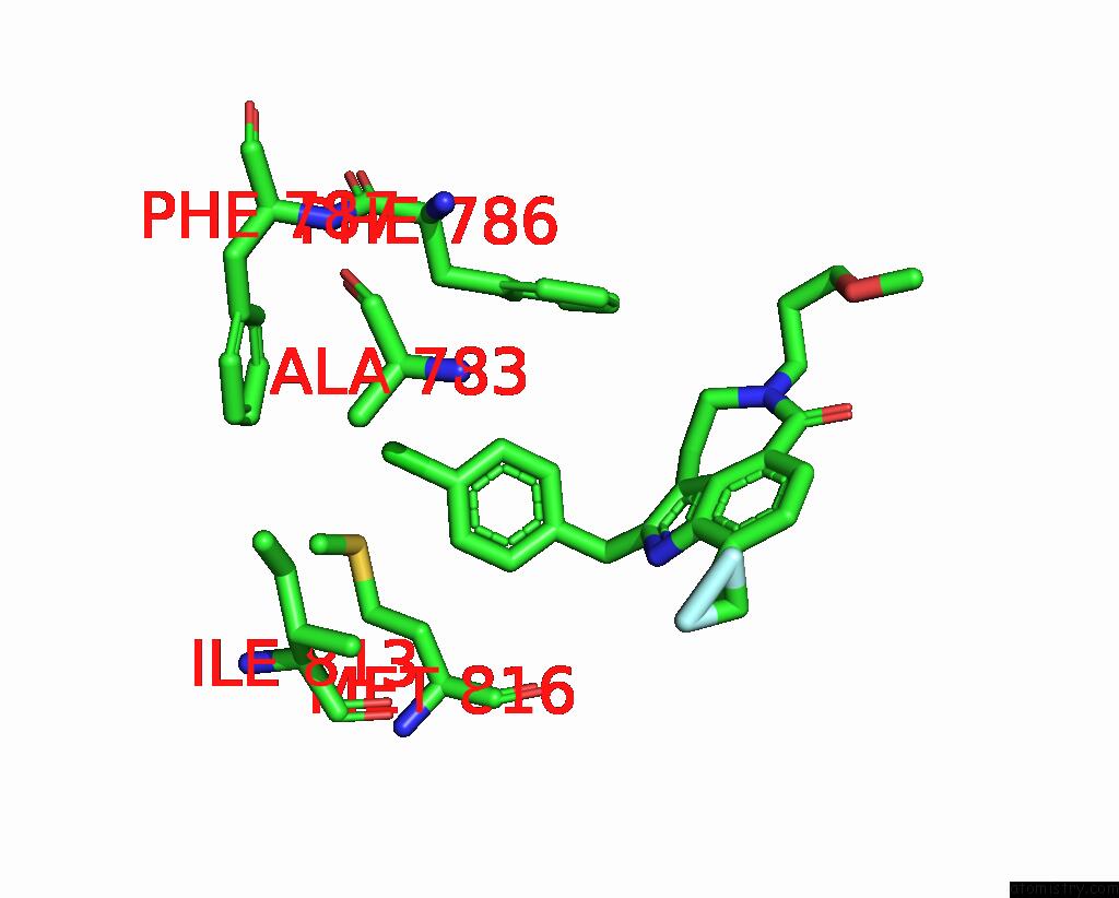
Mono view
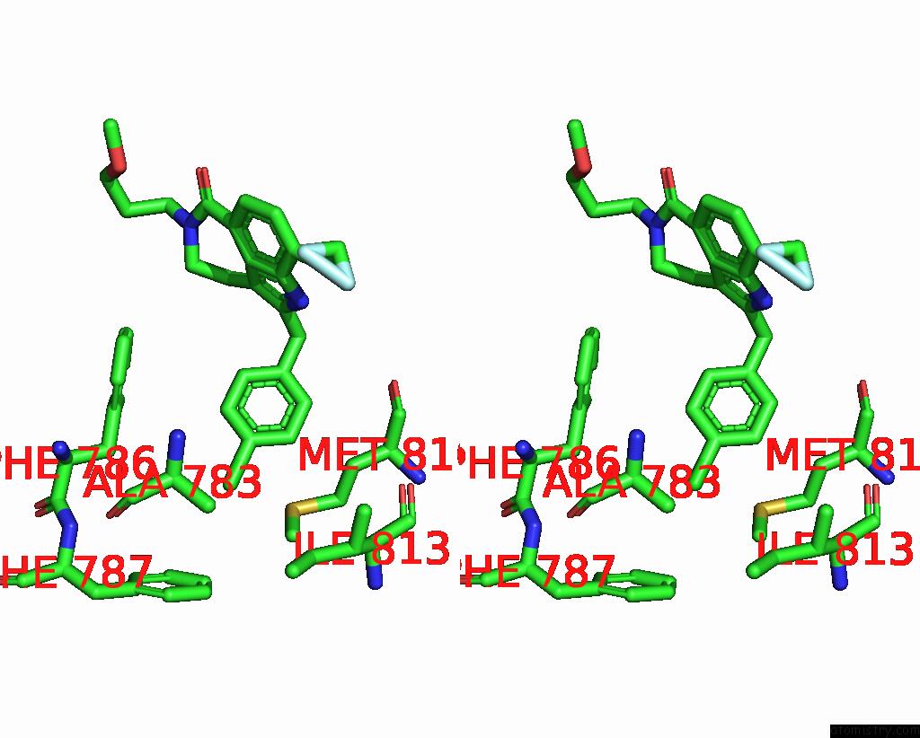
Stereo pair view
Mono view
Stereo pair view
A full contact list of Chlorine with other atoms in the Cl binding
site number 1 of Crystal Structure of PDE5A in Complex with Inhibitor L12 within 5.0Å range:
|
Reference:
D.Wu,
X.Zheng,
R.Liu,
Z.Li,
Z.Jiang,
Q.Zhou,
Y.Huang,
X.N.Wu,
C.Zhang,
Y.Y.Huang,
H.B.Luo.
Free Energy Perturbation (Fep)-Guided Scaffold Hopping. Acta Pharm Sin B V. 12 1351 2022.
ISSN: ISSN 2211-3835
PubMed: 35530128
DOI: 10.1016/J.APSB.2021.09.027
Page generated: Sun Jul 13 00:35:13 2025
ISSN: ISSN 2211-3835
PubMed: 35530128
DOI: 10.1016/J.APSB.2021.09.027
Last articles
Fe in 8Z11Fe in 8Z1W
Fe in 8Z1V
Fe in 8YZ8
Fe in 8YZA
Fe in 8YYP
Fe in 8YXS
Fe in 8YY7
Fe in 8YXT
Fe in 8YKR