Chlorine »
PDB 7fzm-7g2l »
7fzt »
Chlorine in PDB 7fzt: Crystal Structure of Human FABP4 Binding Site Mutated to That of FABP5 in Complex with Rac-(1R,2S)-2-[(3,4-Dichlorobenzoyl) Amino]Cyclohexane-1-Carboxylic Acid
Protein crystallography data
The structure of Crystal Structure of Human FABP4 Binding Site Mutated to That of FABP5 in Complex with Rac-(1R,2S)-2-[(3,4-Dichlorobenzoyl) Amino]Cyclohexane-1-Carboxylic Acid, PDB code: 7fzt
was solved by
A.Ehler,
J.Benz,
U.Obst,
B.Buettelmann,
M.G.Rudolph,
with X-Ray Crystallography technique. A brief refinement statistics is given in the table below:
| Resolution Low / High (Å) | 42.71 / 1.40 |
| Space group | P 2 21 21 |
| Cell size a, b, c (Å), α, β, γ (°) | 32.02, 52.967, 72.208, 90, 90, 90 |
| R / Rfree (%) | 21.5 / 26.5 |
Chlorine Binding Sites:
The binding sites of Chlorine atom in the Crystal Structure of Human FABP4 Binding Site Mutated to That of FABP5 in Complex with Rac-(1R,2S)-2-[(3,4-Dichlorobenzoyl) Amino]Cyclohexane-1-Carboxylic Acid
(pdb code 7fzt). This binding sites where shown within
5.0 Angstroms radius around Chlorine atom.
In total 2 binding sites of Chlorine where determined in the Crystal Structure of Human FABP4 Binding Site Mutated to That of FABP5 in Complex with Rac-(1R,2S)-2-[(3,4-Dichlorobenzoyl) Amino]Cyclohexane-1-Carboxylic Acid, PDB code: 7fzt:
Jump to Chlorine binding site number: 1; 2;
In total 2 binding sites of Chlorine where determined in the Crystal Structure of Human FABP4 Binding Site Mutated to That of FABP5 in Complex with Rac-(1R,2S)-2-[(3,4-Dichlorobenzoyl) Amino]Cyclohexane-1-Carboxylic Acid, PDB code: 7fzt:
Jump to Chlorine binding site number: 1; 2;
Chlorine binding site 1 out of 2 in 7fzt
Go back to
Chlorine binding site 1 out
of 2 in the Crystal Structure of Human FABP4 Binding Site Mutated to That of FABP5 in Complex with Rac-(1R,2S)-2-[(3,4-Dichlorobenzoyl) Amino]Cyclohexane-1-Carboxylic Acid
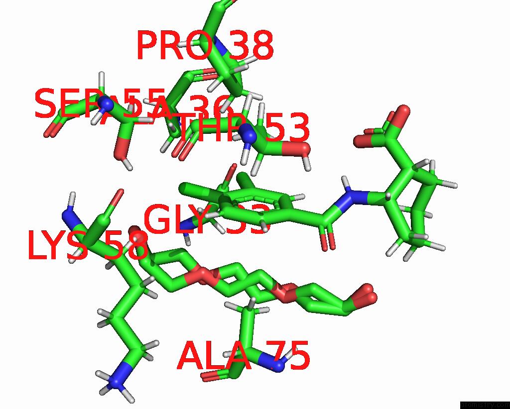
Mono view
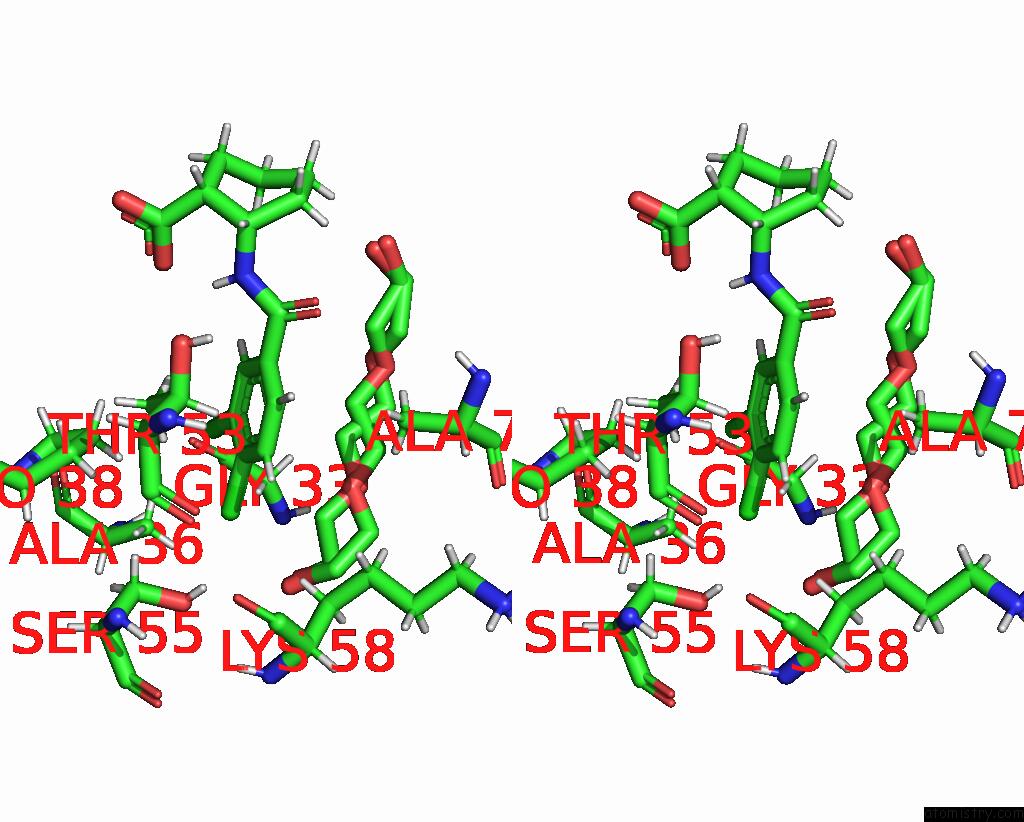
Stereo pair view
Mono view
Stereo pair view
A full contact list of Chlorine with other atoms in the Cl binding
site number 1 of Crystal Structure of Human FABP4 Binding Site Mutated to That of FABP5 in Complex with Rac-(1R,2S)-2-[(3,4-Dichlorobenzoyl) Amino]Cyclohexane-1-Carboxylic Acid within 5.0Å range:
|
Chlorine binding site 2 out of 2 in 7fzt
Go back to
Chlorine binding site 2 out
of 2 in the Crystal Structure of Human FABP4 Binding Site Mutated to That of FABP5 in Complex with Rac-(1R,2S)-2-[(3,4-Dichlorobenzoyl) Amino]Cyclohexane-1-Carboxylic Acid
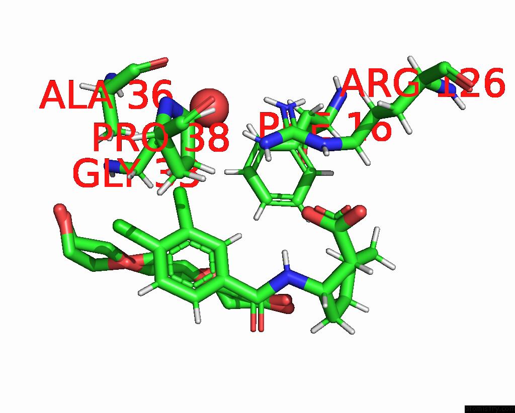
Mono view
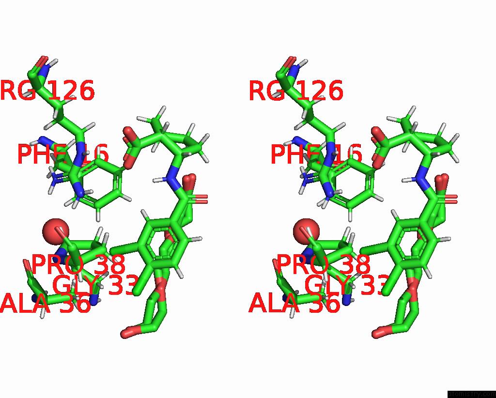
Stereo pair view
Mono view
Stereo pair view
A full contact list of Chlorine with other atoms in the Cl binding
site number 2 of Crystal Structure of Human FABP4 Binding Site Mutated to That of FABP5 in Complex with Rac-(1R,2S)-2-[(3,4-Dichlorobenzoyl) Amino]Cyclohexane-1-Carboxylic Acid within 5.0Å range:
|
Reference:
U.Obst,
C.Magnone,
B.Kuhn,
M.G.Rudolph.
Crystal Structure of A Human FABP4 Binding Site Mutated to That of FABP5 Complex To Be Published.
Page generated: Sun Jul 13 00:57:18 2025
Last articles
K in 8XW9K in 8XTS
K in 8XTR
K in 8XW7
K in 8XW6
K in 8XTQ
K in 8XMI
K in 8XTP
K in 8XMH
K in 8X9Q