Chlorine »
PDB 7guh-7h7i »
7guh »
Chlorine in PDB 7guh: Crystal Structure of B-Cell Lymphoma 6 Protein Btb Domain in Complex with Ligand 1 at 7.55 Mgy X-Ray Dose.
Protein crystallography data
The structure of Crystal Structure of B-Cell Lymphoma 6 Protein Btb Domain in Complex with Ligand 1 at 7.55 Mgy X-Ray Dose., PDB code: 7guh
was solved by
M.J.Rodrigues,
Y.V.Le Bihan,
R.L.M.Van Montfort,
with X-Ray Crystallography technique. A brief refinement statistics is given in the table below:
| Resolution Low / High (Å) | 33.88 / 1.80 |
| Space group | P 61 2 2 |
| Cell size a, b, c (Å), α, β, γ (°) | 67.55, 67.55, 166.22, 90, 90, 120 |
| R / Rfree (%) | 17.8 / 20.1 |
Chlorine Binding Sites:
The binding sites of Chlorine atom in the Crystal Structure of B-Cell Lymphoma 6 Protein Btb Domain in Complex with Ligand 1 at 7.55 Mgy X-Ray Dose.
(pdb code 7guh). This binding sites where shown within
5.0 Angstroms radius around Chlorine atom.
In total 2 binding sites of Chlorine where determined in the Crystal Structure of B-Cell Lymphoma 6 Protein Btb Domain in Complex with Ligand 1 at 7.55 Mgy X-Ray Dose., PDB code: 7guh:
Jump to Chlorine binding site number: 1; 2;
In total 2 binding sites of Chlorine where determined in the Crystal Structure of B-Cell Lymphoma 6 Protein Btb Domain in Complex with Ligand 1 at 7.55 Mgy X-Ray Dose., PDB code: 7guh:
Jump to Chlorine binding site number: 1; 2;
Chlorine binding site 1 out of 2 in 7guh
Go back to
Chlorine binding site 1 out
of 2 in the Crystal Structure of B-Cell Lymphoma 6 Protein Btb Domain in Complex with Ligand 1 at 7.55 Mgy X-Ray Dose.
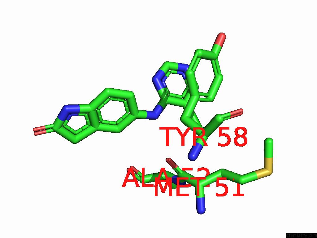
Mono view
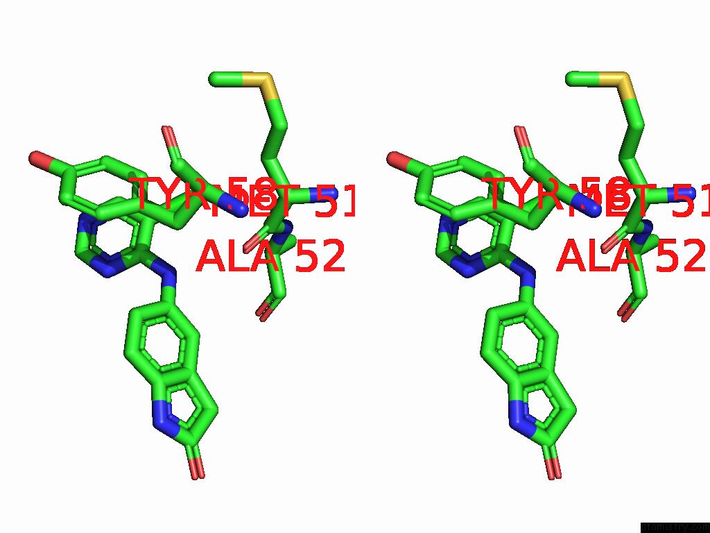
Stereo pair view
Mono view
Stereo pair view
A full contact list of Chlorine with other atoms in the Cl binding
site number 1 of Crystal Structure of B-Cell Lymphoma 6 Protein Btb Domain in Complex with Ligand 1 at 7.55 Mgy X-Ray Dose. within 5.0Å range:
|
Chlorine binding site 2 out of 2 in 7guh
Go back to
Chlorine binding site 2 out
of 2 in the Crystal Structure of B-Cell Lymphoma 6 Protein Btb Domain in Complex with Ligand 1 at 7.55 Mgy X-Ray Dose.
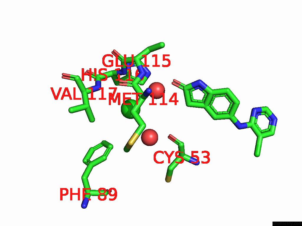
Mono view
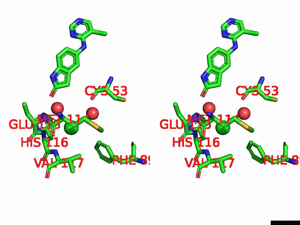
Stereo pair view
Mono view
Stereo pair view
A full contact list of Chlorine with other atoms in the Cl binding
site number 2 of Crystal Structure of B-Cell Lymphoma 6 Protein Btb Domain in Complex with Ligand 1 at 7.55 Mgy X-Ray Dose. within 5.0Å range:
|
Reference:
M.J.Rodrigues,
M.Cabry,
G.Collie,
M.Carter,
C.Mcandrew,
R.L.Owen,
B.R.Bellenie,
Y.V.Le Bihan,
R.L.M.Van Montfort.
Specific Radiation Damage to Halogenated Inhibitors and Ligands in Protein-Ligand Crystal Structures. J.Appl.Crystallogr. V. 57 1951 2024.
ISSN: ISSN 0021-8898
PubMed: 39628887
DOI: 10.1107/S1600576724010549
Page generated: Sun Jul 13 02:07:16 2025
ISSN: ISSN 0021-8898
PubMed: 39628887
DOI: 10.1107/S1600576724010549
Last articles
Na in 5IHGNa in 5IHF
Na in 5IER
Na in 5IF6
Na in 5IDD
Na in 5ICM
Na in 5ICC
Na in 5ICF
Na in 5IBC
Na in 5IAS