Chlorine »
PDB 7l9j-7lgp »
7l9u »
Chlorine in PDB 7l9u: Crystal Structure of S-Adenosylmethionine-Dependent Methyltransferase Umaa From Mycobacterium Tuberculosis in Complex with A 12-Mer Peg
Protein crystallography data
The structure of Crystal Structure of S-Adenosylmethionine-Dependent Methyltransferase Umaa From Mycobacterium Tuberculosis in Complex with A 12-Mer Peg, PDB code: 7l9u
was solved by
Seattle Structural Genomics Center For Infectious Disease,
Seattlestructural Genomics Center For Infectious Disease (Ssgcid),
with X-Ray Crystallography technique. A brief refinement statistics is given in the table below:
| Resolution Low / High (Å) | 43.46 / 1.55 |
| Space group | P 32 2 1 |
| Cell size a, b, c (Å), α, β, γ (°) | 103.02, 103.02, 49.76, 90, 90, 120 |
| R / Rfree (%) | 15.7 / 17.5 |
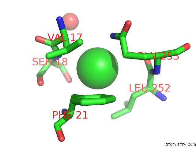
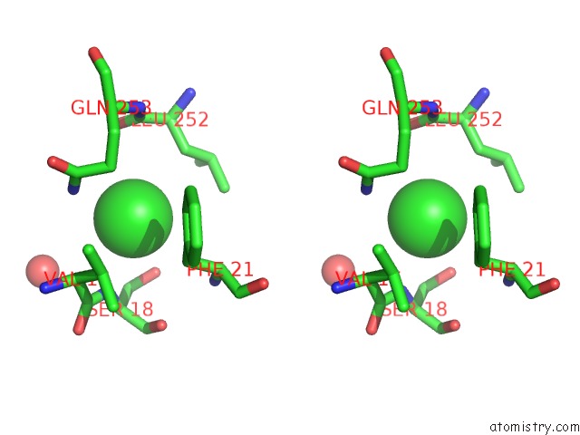
Chlorine Binding Sites:
The binding sites of Chlorine atom in the Crystal Structure of S-Adenosylmethionine-Dependent Methyltransferase Umaa From Mycobacterium Tuberculosis in Complex with A 12-Mer Peg
(pdb code 7l9u). This binding sites where shown within
5.0 Angstroms radius around Chlorine atom.
In total only one binding site of Chlorine was determined in the Crystal Structure of S-Adenosylmethionine-Dependent Methyltransferase Umaa From Mycobacterium Tuberculosis in Complex with A 12-Mer Peg, PDB code: 7l9u:
In total only one binding site of Chlorine was determined in the Crystal Structure of S-Adenosylmethionine-Dependent Methyltransferase Umaa From Mycobacterium Tuberculosis in Complex with A 12-Mer Peg, PDB code: 7l9u:
Chlorine binding site 1 out of 1 in 7l9u
Go back to
Chlorine binding site 1 out
of 1 in the Crystal Structure of S-Adenosylmethionine-Dependent Methyltransferase Umaa From Mycobacterium Tuberculosis in Complex with A 12-Mer Peg
Mono view
Stereo pair view
Mono view
Stereo pair view
A full contact list of Chlorine with other atoms in the Cl binding
site number 1 of Crystal Structure of S-Adenosylmethionine-Dependent Methyltransferase Umaa From Mycobacterium Tuberculosis in Complex with A 12-Mer Peg within 5.0Å range:
|
Reference:
J.Abendroth,
M.J.Weiss,
D.D.Lorimer,
P.S.Horanyi,
T.E.Edwards.
Crystal Structure of S-Adenosylmethionine-Dependent Methyltransferase Umaa From Mycobacterium Tuberculosis in Complex with A 12-Mer Peg To Be Published.
Page generated: Sun Jul 13 03:38:23 2025
Last articles
Fe in 2FRFFe in 2FRC
Fe in 2FR7
Fe in 2FLA
Fe in 2FNQ
Fe in 2FMY
Fe in 2FLQ
Fe in 2FKZ
Fe in 2FJE
Fe in 2FJD