Chlorine »
PDB 7n14-7neu »
7n63 »
Chlorine in PDB 7n63: X-Ray Structure of HCAN_0200, An Aminotransferase From Helicobacter Canadensis in Complex with Its External Aldimine
Protein crystallography data
The structure of X-Ray Structure of HCAN_0200, An Aminotransferase From Helicobacter Canadensis in Complex with Its External Aldimine, PDB code: 7n63
was solved by
W.A.Griffiths,
C.J.Heisdorf,
J.B.Thoden,
H.M.Holden,
with X-Ray Crystallography technique. A brief refinement statistics is given in the table below:
| Resolution Low / High (Å) | 35.71 / 1.40 |
| Space group | C 2 2 21 |
| Cell size a, b, c (Å), α, β, γ (°) | 61.005, 150.766, 91.058, 90, 90, 90 |
| R / Rfree (%) | 17.1 / 19 |
Other elements in 7n63:
The structure of X-Ray Structure of HCAN_0200, An Aminotransferase From Helicobacter Canadensis in Complex with Its External Aldimine also contains other interesting chemical elements:
| Magnesium | (Mg) | 1 atom |
Chlorine Binding Sites:
The binding sites of Chlorine atom in the X-Ray Structure of HCAN_0200, An Aminotransferase From Helicobacter Canadensis in Complex with Its External Aldimine
(pdb code 7n63). This binding sites where shown within
5.0 Angstroms radius around Chlorine atom.
In total only one binding site of Chlorine was determined in the X-Ray Structure of HCAN_0200, An Aminotransferase From Helicobacter Canadensis in Complex with Its External Aldimine, PDB code: 7n63:
In total only one binding site of Chlorine was determined in the X-Ray Structure of HCAN_0200, An Aminotransferase From Helicobacter Canadensis in Complex with Its External Aldimine, PDB code: 7n63:
Chlorine binding site 1 out of 1 in 7n63
Go back to
Chlorine binding site 1 out
of 1 in the X-Ray Structure of HCAN_0200, An Aminotransferase From Helicobacter Canadensis in Complex with Its External Aldimine
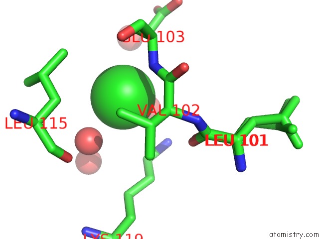
Mono view
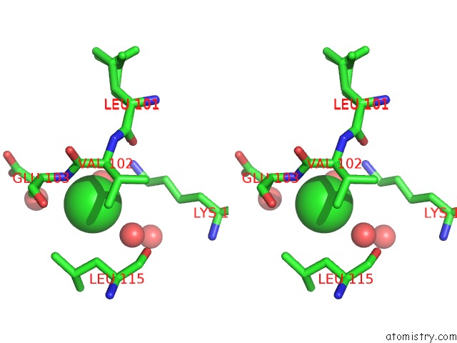
Stereo pair view
Mono view
Stereo pair view
A full contact list of Chlorine with other atoms in the Cl binding
site number 1 of X-Ray Structure of HCAN_0200, An Aminotransferase From Helicobacter Canadensis in Complex with Its External Aldimine within 5.0Å range:
|
Reference:
C.J.Heisdorf,
W.A.Griffiths,
J.B.Thoden,
H.M.Holden.
Investigation of the Enzymes Required For the Biosynthesis of An Unusual Formylated Sugar in the Emerging Human Pathogen Helicobacter Canadensis To Be Published.
Page generated: Sun Jul 13 04:19:10 2025
Last articles
Mg in 6VZSMg in 6VZT
Mg in 6VZR
Mg in 6VZQ
Mg in 6VZ8
Mg in 6VZ9
Mg in 6VYD
Mg in 6VZ4
Mg in 6VWP
Mg in 6VWW