Chlorine »
PDB 7o3p-7o6j »
7o5m »
Chlorine in PDB 7o5m: Crystal Structure of S-Adenosyl-L-Homocysteine Hydrolase From Synechocystis Sp. Pcc 6803 Cocrystallized with Adenosine in the Presence of Na+ Cations
Enzymatic activity of Crystal Structure of S-Adenosyl-L-Homocysteine Hydrolase From Synechocystis Sp. Pcc 6803 Cocrystallized with Adenosine in the Presence of Na+ Cations
All present enzymatic activity of Crystal Structure of S-Adenosyl-L-Homocysteine Hydrolase From Synechocystis Sp. Pcc 6803 Cocrystallized with Adenosine in the Presence of Na+ Cations:
3.3.1.1;
3.3.1.1;
Protein crystallography data
The structure of Crystal Structure of S-Adenosyl-L-Homocysteine Hydrolase From Synechocystis Sp. Pcc 6803 Cocrystallized with Adenosine in the Presence of Na+ Cations, PDB code: 7o5m
was solved by
P.H.Malecki,
B.Imiolczyk,
J.Barciszewski,
J.Czyrko-Horczak,
K.Brzezinski,
with X-Ray Crystallography technique. A brief refinement statistics is given in the table below:
| Resolution Low / High (Å) | 42.27 / 2.20 |
| Space group | C 2 2 21 |
| Cell size a, b, c (Å), α, β, γ (°) | 120.77, 197.13, 82.23, 90, 90, 90 |
| R / Rfree (%) | 19.2 / 24.9 |
Chlorine Binding Sites:
The binding sites of Chlorine atom in the Crystal Structure of S-Adenosyl-L-Homocysteine Hydrolase From Synechocystis Sp. Pcc 6803 Cocrystallized with Adenosine in the Presence of Na+ Cations
(pdb code 7o5m). This binding sites where shown within
5.0 Angstroms radius around Chlorine atom.
In total 3 binding sites of Chlorine where determined in the Crystal Structure of S-Adenosyl-L-Homocysteine Hydrolase From Synechocystis Sp. Pcc 6803 Cocrystallized with Adenosine in the Presence of Na+ Cations, PDB code: 7o5m:
Jump to Chlorine binding site number: 1; 2; 3;
In total 3 binding sites of Chlorine where determined in the Crystal Structure of S-Adenosyl-L-Homocysteine Hydrolase From Synechocystis Sp. Pcc 6803 Cocrystallized with Adenosine in the Presence of Na+ Cations, PDB code: 7o5m:
Jump to Chlorine binding site number: 1; 2; 3;
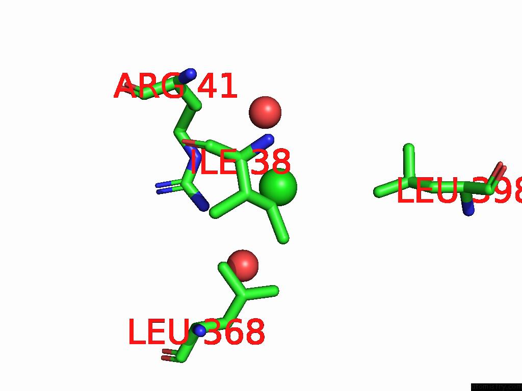
Chlorine binding site 1 out of 3 in 7o5m
Go back to
Chlorine binding site 1 out
of 3 in the Crystal Structure of S-Adenosyl-L-Homocysteine Hydrolase From Synechocystis Sp. Pcc 6803 Cocrystallized with Adenosine in the Presence of Na+ Cations
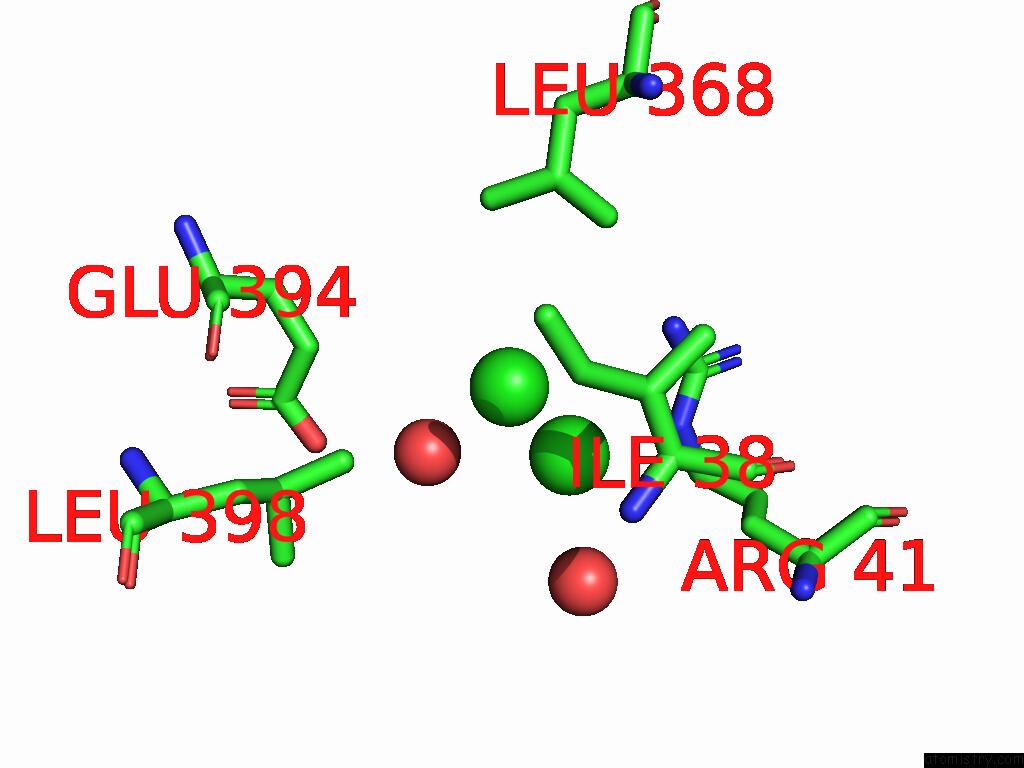
Mono view
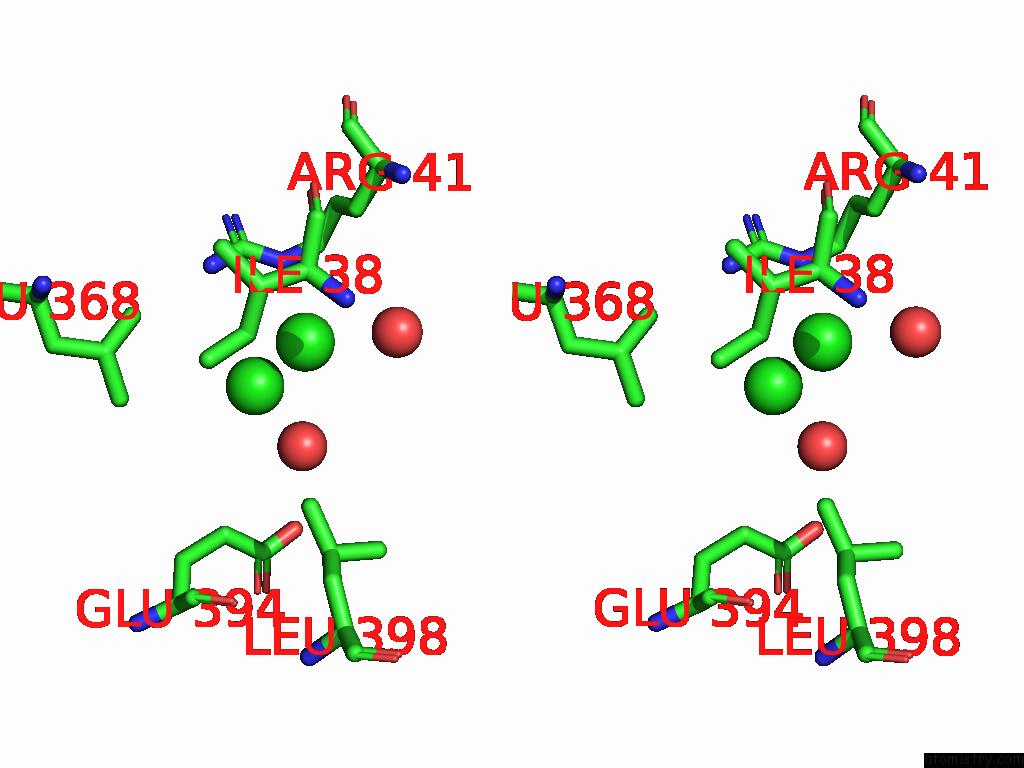
Stereo pair view
Mono view
Stereo pair view
A full contact list of Chlorine with other atoms in the Cl binding
site number 1 of Crystal Structure of S-Adenosyl-L-Homocysteine Hydrolase From Synechocystis Sp. Pcc 6803 Cocrystallized with Adenosine in the Presence of Na+ Cations within 5.0Å range:
|
Chlorine binding site 2 out of 3 in 7o5m
Go back to
Chlorine binding site 2 out
of 3 in the Crystal Structure of S-Adenosyl-L-Homocysteine Hydrolase From Synechocystis Sp. Pcc 6803 Cocrystallized with Adenosine in the Presence of Na+ Cations
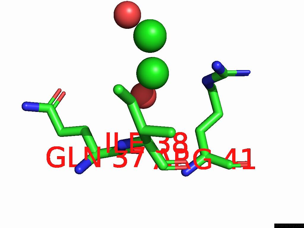
Mono view
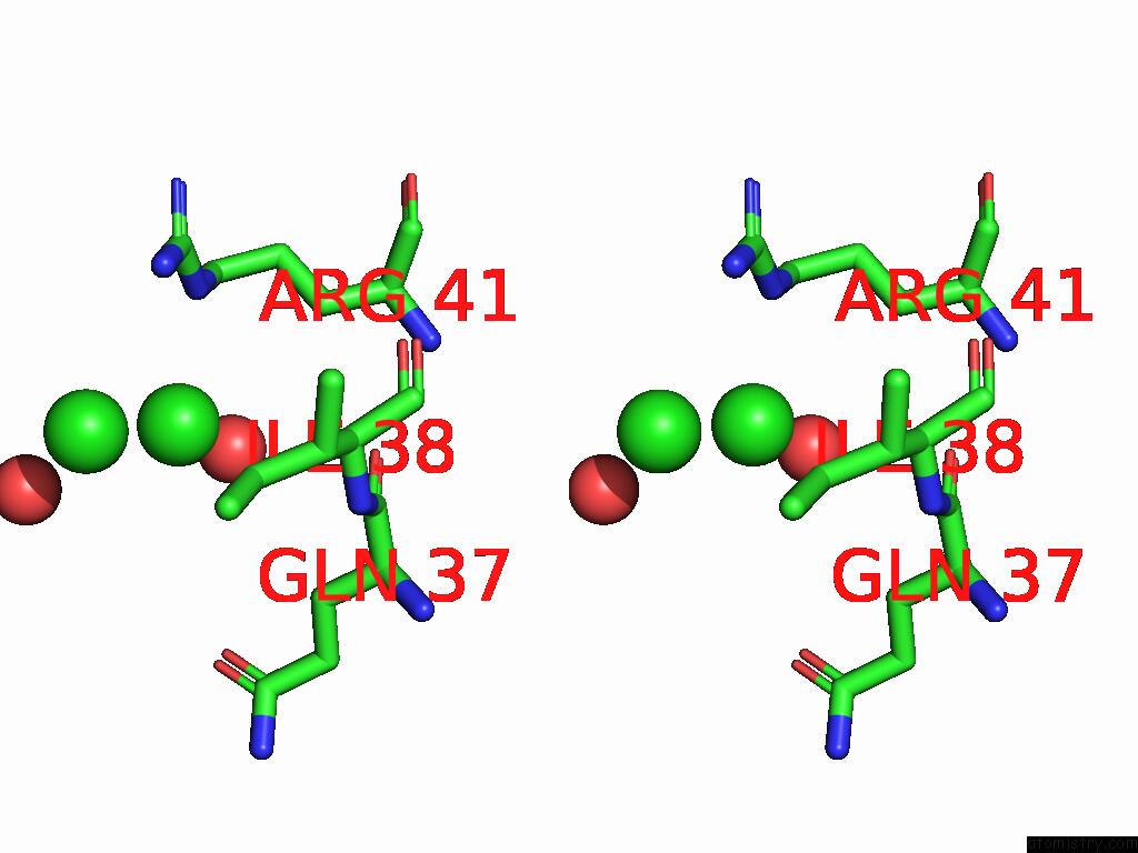
Stereo pair view
Mono view
Stereo pair view
A full contact list of Chlorine with other atoms in the Cl binding
site number 2 of Crystal Structure of S-Adenosyl-L-Homocysteine Hydrolase From Synechocystis Sp. Pcc 6803 Cocrystallized with Adenosine in the Presence of Na+ Cations within 5.0Å range:
|
Chlorine binding site 3 out of 3 in 7o5m
Go back to
Chlorine binding site 3 out
of 3 in the Crystal Structure of S-Adenosyl-L-Homocysteine Hydrolase From Synechocystis Sp. Pcc 6803 Cocrystallized with Adenosine in the Presence of Na+ Cations
Mono view

Stereo pair view
Mono view
Stereo pair view
A full contact list of Chlorine with other atoms in the Cl binding
site number 3 of Crystal Structure of S-Adenosyl-L-Homocysteine Hydrolase From Synechocystis Sp. Pcc 6803 Cocrystallized with Adenosine in the Presence of Na+ Cations within 5.0Å range:
|
Reference:
P.H.Malecki,
B.Imiolczyk,
J.Barciszewski,
J.Czyrko-Horczak,
J.Sliwiak,
M.Gawel,
K.Wozniak,
M.Jaskolski,
K.Brzezinski.
Biochemical and Structural Insights Into An Unusual, Alkali-Metal-Independent S-Adenosyl-L-Homocysteine Hydrolase From Synechocystis Sp. Pcc 6803. Acta Crystallogr D Struct V. 78 865 2022BIOL.
ISSN: ISSN 2059-7983
PubMed: 35775986
DOI: 10.1107/S2059798322005605
Page generated: Sun Jul 13 04:43:08 2025
ISSN: ISSN 2059-7983
PubMed: 35775986
DOI: 10.1107/S2059798322005605
Last articles
Fe in 9ERLFe in 9ERK
Fe in 9ERJ
Fe in 9ERI
Fe in 9ERB
Fe in 9ERC
Fe in 9ER7
Fe in 9ERA
Fe in 9ER6
Fe in 9ER5