Chlorine »
PDB 7o3p-7o6j »
7o66 »
Chlorine in PDB 7o66: Crystal Structure of Human Mitochondrial Ferritin (Hmtf) Fe(II)-Loaded For 60 Minutes Showing Either A Dioxygen or A Superoxide Anion Coordinated to Iron Ions in the Ferroxidase Site
Enzymatic activity of Crystal Structure of Human Mitochondrial Ferritin (Hmtf) Fe(II)-Loaded For 60 Minutes Showing Either A Dioxygen or A Superoxide Anion Coordinated to Iron Ions in the Ferroxidase Site
All present enzymatic activity of Crystal Structure of Human Mitochondrial Ferritin (Hmtf) Fe(II)-Loaded For 60 Minutes Showing Either A Dioxygen or A Superoxide Anion Coordinated to Iron Ions in the Ferroxidase Site:
1.16.3.1;
1.16.3.1;
Protein crystallography data
The structure of Crystal Structure of Human Mitochondrial Ferritin (Hmtf) Fe(II)-Loaded For 60 Minutes Showing Either A Dioxygen or A Superoxide Anion Coordinated to Iron Ions in the Ferroxidase Site, PDB code: 7o66
was solved by
C.Pozzi,
S.Ciambellotti,
G.Tassone,
P.Turano,
S.Mangani,
with X-Ray Crystallography technique. A brief refinement statistics is given in the table below:
| Resolution Low / High (Å) | 64.99 / 1.60 |
| Space group | F 4 3 2 |
| Cell size a, b, c (Å), α, β, γ (°) | 183.83, 183.83, 183.83, 90, 90, 90 |
| R / Rfree (%) | 15.5 / 17.9 |
Other elements in 7o66:
The structure of Crystal Structure of Human Mitochondrial Ferritin (Hmtf) Fe(II)-Loaded For 60 Minutes Showing Either A Dioxygen or A Superoxide Anion Coordinated to Iron Ions in the Ferroxidase Site also contains other interesting chemical elements:
| Iron | (Fe) | 11 atoms |
| Magnesium | (Mg) | 1 atom |
Chlorine Binding Sites:
The binding sites of Chlorine atom in the Crystal Structure of Human Mitochondrial Ferritin (Hmtf) Fe(II)-Loaded For 60 Minutes Showing Either A Dioxygen or A Superoxide Anion Coordinated to Iron Ions in the Ferroxidase Site
(pdb code 7o66). This binding sites where shown within
5.0 Angstroms radius around Chlorine atom.
In total 6 binding sites of Chlorine where determined in the Crystal Structure of Human Mitochondrial Ferritin (Hmtf) Fe(II)-Loaded For 60 Minutes Showing Either A Dioxygen or A Superoxide Anion Coordinated to Iron Ions in the Ferroxidase Site, PDB code: 7o66:
Jump to Chlorine binding site number: 1; 2; 3; 4; 5; 6;
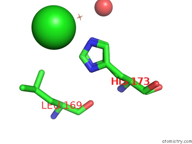
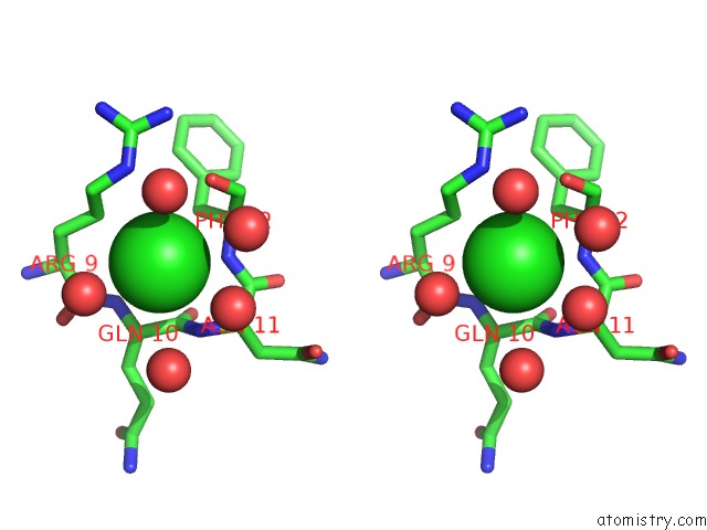
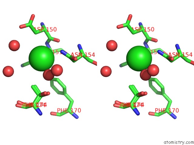
Chlorine binding site 1 out of 6 in 7o66
Go back to
Chlorine binding site 1 out
of 6 in the Crystal Structure of Human Mitochondrial Ferritin (Hmtf) Fe(II)-Loaded For 60 Minutes Showing Either A Dioxygen or A Superoxide Anion Coordinated to Iron Ions in the Ferroxidase Site
Mono view

Stereo pair view
Mono view
Stereo pair view
A full contact list of Chlorine with other atoms in the Cl binding
site number 1 of Crystal Structure of Human Mitochondrial Ferritin (Hmtf) Fe(II)-Loaded For 60 Minutes Showing Either A Dioxygen or A Superoxide Anion Coordinated to Iron Ions in the Ferroxidase Site within 5.0Å range:
|
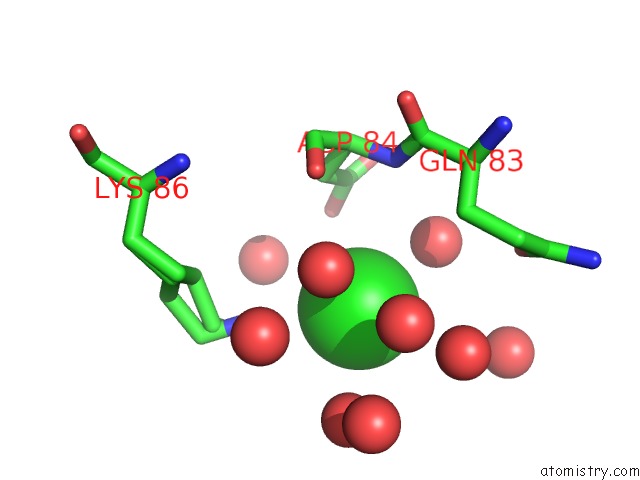
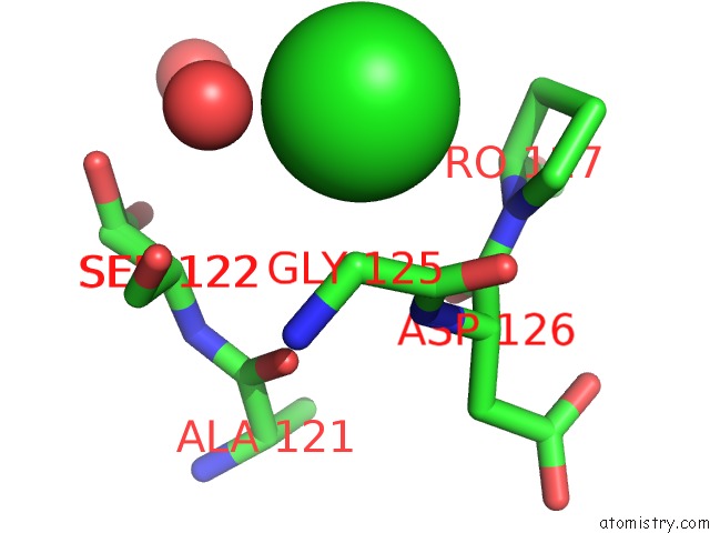
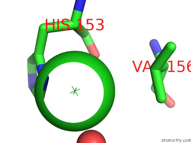
Chlorine binding site 2 out of 6 in 7o66
Go back to
Chlorine binding site 2 out
of 6 in the Crystal Structure of Human Mitochondrial Ferritin (Hmtf) Fe(II)-Loaded For 60 Minutes Showing Either A Dioxygen or A Superoxide Anion Coordinated to Iron Ions in the Ferroxidase Site
Mono view
Stereo pair view
Mono view
Stereo pair view
A full contact list of Chlorine with other atoms in the Cl binding
site number 2 of Crystal Structure of Human Mitochondrial Ferritin (Hmtf) Fe(II)-Loaded For 60 Minutes Showing Either A Dioxygen or A Superoxide Anion Coordinated to Iron Ions in the Ferroxidase Site within 5.0Å range:
|
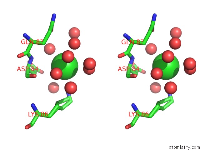
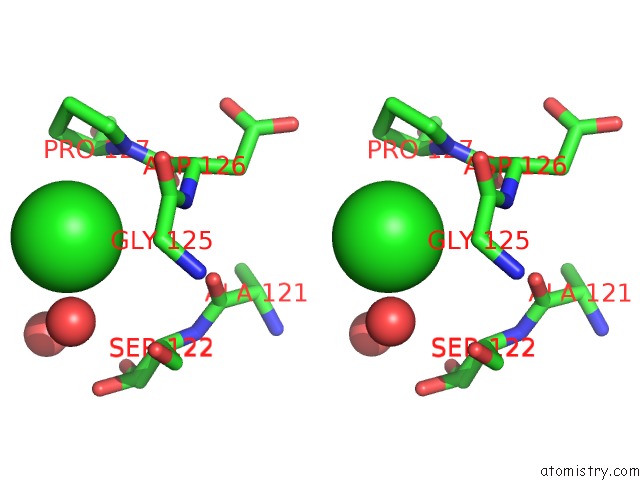
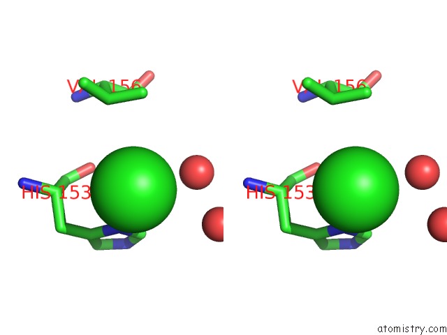
Chlorine binding site 3 out of 6 in 7o66
Go back to
Chlorine binding site 3 out
of 6 in the Crystal Structure of Human Mitochondrial Ferritin (Hmtf) Fe(II)-Loaded For 60 Minutes Showing Either A Dioxygen or A Superoxide Anion Coordinated to Iron Ions in the Ferroxidase Site
Mono view
Stereo pair view
Mono view
Stereo pair view
A full contact list of Chlorine with other atoms in the Cl binding
site number 3 of Crystal Structure of Human Mitochondrial Ferritin (Hmtf) Fe(II)-Loaded For 60 Minutes Showing Either A Dioxygen or A Superoxide Anion Coordinated to Iron Ions in the Ferroxidase Site within 5.0Å range:
|
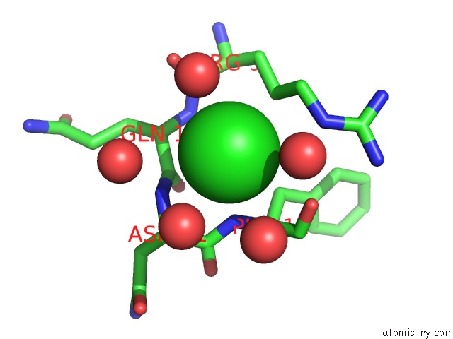
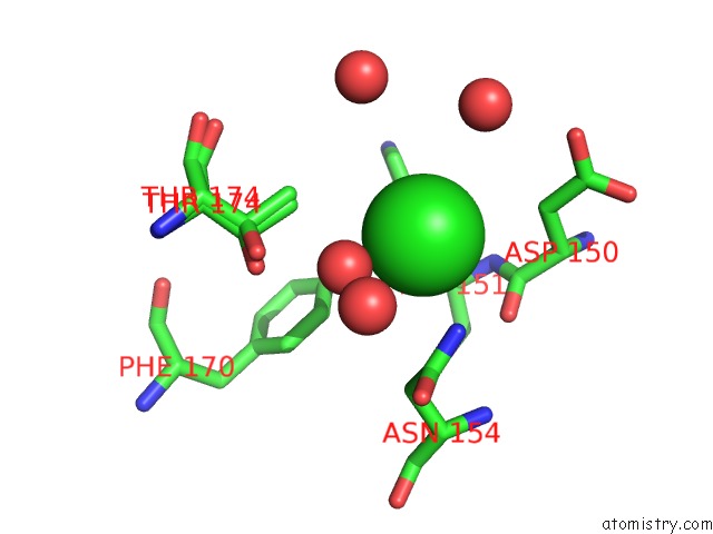
Chlorine binding site 4 out of 6 in 7o66
Go back to
Chlorine binding site 4 out
of 6 in the Crystal Structure of Human Mitochondrial Ferritin (Hmtf) Fe(II)-Loaded For 60 Minutes Showing Either A Dioxygen or A Superoxide Anion Coordinated to Iron Ions in the Ferroxidase Site
Mono view
Stereo pair view
Mono view
Stereo pair view
A full contact list of Chlorine with other atoms in the Cl binding
site number 4 of Crystal Structure of Human Mitochondrial Ferritin (Hmtf) Fe(II)-Loaded For 60 Minutes Showing Either A Dioxygen or A Superoxide Anion Coordinated to Iron Ions in the Ferroxidase Site within 5.0Å range:
|
Chlorine binding site 5 out of 6 in 7o66
Go back to
Chlorine binding site 5 out
of 6 in the Crystal Structure of Human Mitochondrial Ferritin (Hmtf) Fe(II)-Loaded For 60 Minutes Showing Either A Dioxygen or A Superoxide Anion Coordinated to Iron Ions in the Ferroxidase Site
Mono view
Stereo pair view
Mono view
Stereo pair view
A full contact list of Chlorine with other atoms in the Cl binding
site number 5 of Crystal Structure of Human Mitochondrial Ferritin (Hmtf) Fe(II)-Loaded For 60 Minutes Showing Either A Dioxygen or A Superoxide Anion Coordinated to Iron Ions in the Ferroxidase Site within 5.0Å range:
|
Chlorine binding site 6 out of 6 in 7o66
Go back to
Chlorine binding site 6 out
of 6 in the Crystal Structure of Human Mitochondrial Ferritin (Hmtf) Fe(II)-Loaded For 60 Minutes Showing Either A Dioxygen or A Superoxide Anion Coordinated to Iron Ions in the Ferroxidase Site
Mono view
Stereo pair view
Mono view
Stereo pair view
A full contact list of Chlorine with other atoms in the Cl binding
site number 6 of Crystal Structure of Human Mitochondrial Ferritin (Hmtf) Fe(II)-Loaded For 60 Minutes Showing Either A Dioxygen or A Superoxide Anion Coordinated to Iron Ions in the Ferroxidase Site within 5.0Å range:
|
Reference:
S.Ciambellotti,
A.Pratesi,
G.Tassone,
P.Turano,
S.Mangani,
C.Pozzi.
Iron Binding in the Ferroxidase Site of Human Mitochondrial Ferritin. Chemistry 2021.
ISSN: ISSN 0947-6539
PubMed: 34343376
DOI: 10.1002/CHEM.202102270
Page generated: Sun Jul 13 04:44:11 2025
ISSN: ISSN 0947-6539
PubMed: 34343376
DOI: 10.1002/CHEM.202102270
Last articles
Mg in 6KF3Mg in 6KE4
Mg in 6KE0
Mg in 6KDZ
Mg in 6KDX
Mg in 6KDN
Mg in 6KDM
Mg in 6KDK
Mg in 6KDJ
Mg in 6KD2