Chlorine »
PDB 7p2t-7pe0 »
7p2v »
Chlorine in PDB 7p2v: Crystal Structure of Schistosoma Mansoni HDAC8 in Complex with A 4- Chlorophenyl-Spiroindoline Capped Hydroxamate-Based Inhibitor, Bound to A Novel Site
Protein crystallography data
The structure of Crystal Structure of Schistosoma Mansoni HDAC8 in Complex with A 4- Chlorophenyl-Spiroindoline Capped Hydroxamate-Based Inhibitor, Bound to A Novel Site, PDB code: 7p2v
was solved by
F.Saccoccia,
S.Gemma,
G.Campiani,
G.Ruberti,
with X-Ray Crystallography technique. A brief refinement statistics is given in the table below:
| Resolution Low / High (Å) | 39.14 / 2.25 |
| Space group | P 41 21 2 |
| Cell size a, b, c (Å), α, β, γ (°) | 71.14, 71.14, 186.911, 90, 90, 90 |
| R / Rfree (%) | 21.2 / 25.2 |
Other elements in 7p2v:
The structure of Crystal Structure of Schistosoma Mansoni HDAC8 in Complex with A 4- Chlorophenyl-Spiroindoline Capped Hydroxamate-Based Inhibitor, Bound to A Novel Site also contains other interesting chemical elements:
| Zinc | (Zn) | 1 atom |
| Potassium | (K) | 2 atoms |
Chlorine Binding Sites:
The binding sites of Chlorine atom in the Crystal Structure of Schistosoma Mansoni HDAC8 in Complex with A 4- Chlorophenyl-Spiroindoline Capped Hydroxamate-Based Inhibitor, Bound to A Novel Site
(pdb code 7p2v). This binding sites where shown within
5.0 Angstroms radius around Chlorine atom.
In total only one binding site of Chlorine was determined in the Crystal Structure of Schistosoma Mansoni HDAC8 in Complex with A 4- Chlorophenyl-Spiroindoline Capped Hydroxamate-Based Inhibitor, Bound to A Novel Site, PDB code: 7p2v:
In total only one binding site of Chlorine was determined in the Crystal Structure of Schistosoma Mansoni HDAC8 in Complex with A 4- Chlorophenyl-Spiroindoline Capped Hydroxamate-Based Inhibitor, Bound to A Novel Site, PDB code: 7p2v:
Chlorine binding site 1 out of 1 in 7p2v
Go back to
Chlorine binding site 1 out
of 1 in the Crystal Structure of Schistosoma Mansoni HDAC8 in Complex with A 4- Chlorophenyl-Spiroindoline Capped Hydroxamate-Based Inhibitor, Bound to A Novel Site
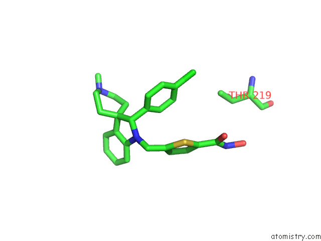
Mono view
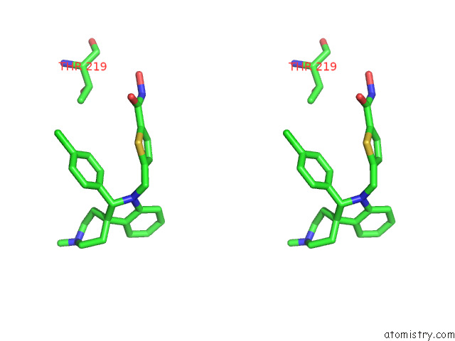
Stereo pair view
Mono view
Stereo pair view
A full contact list of Chlorine with other atoms in the Cl binding
site number 1 of Crystal Structure of Schistosoma Mansoni HDAC8 in Complex with A 4- Chlorophenyl-Spiroindoline Capped Hydroxamate-Based Inhibitor, Bound to A Novel Site within 5.0Å range:
|
Reference:
F.Saccoccia,
L.Pozzetti,
R.Gimmelli,
S.Butini,
A.Guidi,
G.Papoff,
M.Giannaccari,
S.Brogi,
V.Scognamiglio,
S.Gemma,
G.Ruberti,
G.Campiani.
Crystal Structures of Schistosoma Mansoni Histone Deacetylase 8 Reveal A Novel Binding Site For Allosteric Inhibitors. J.Biol.Chem. V. 298 02375 2022.
ISSN: ESSN 1083-351X
PubMed: 35970392
DOI: 10.1016/J.JBC.2022.102375
Page generated: Sun Jul 13 05:32:35 2025
ISSN: ESSN 1083-351X
PubMed: 35970392
DOI: 10.1016/J.JBC.2022.102375
Last articles
Fe in 2YXOFe in 2YRS
Fe in 2YXC
Fe in 2YNM
Fe in 2YVJ
Fe in 2YP1
Fe in 2YU2
Fe in 2YU1
Fe in 2YQB
Fe in 2YOO