Chlorine »
PDB 7pow-7pvn »
7ppu »
Chlorine in PDB 7ppu: Structure of Dife-Sulerythrin at 0.57 Mgy Total Absorbed Dose
Protein crystallography data
The structure of Structure of Dife-Sulerythrin at 0.57 Mgy Total Absorbed Dose, PDB code: 7ppu
was solved by
F.Lennartz,
M.S.Weiss,
with X-Ray Crystallography technique. A brief refinement statistics is given in the table below:
| Resolution Low / High (Å) | 38.53 / 1.34 |
| Space group | P 63 |
| Cell size a, b, c (Å), α, β, γ (°) | 72.02, 72.02, 97.997, 90, 90, 120 |
| R / Rfree (%) | 18.5 / 21.5 |
Other elements in 7ppu:
The structure of Structure of Dife-Sulerythrin at 0.57 Mgy Total Absorbed Dose also contains other interesting chemical elements:
| Iron | (Fe) | 4 atoms |
Chlorine Binding Sites:
The binding sites of Chlorine atom in the Structure of Dife-Sulerythrin at 0.57 Mgy Total Absorbed Dose
(pdb code 7ppu). This binding sites where shown within
5.0 Angstroms radius around Chlorine atom.
In total 2 binding sites of Chlorine where determined in the Structure of Dife-Sulerythrin at 0.57 Mgy Total Absorbed Dose, PDB code: 7ppu:
Jump to Chlorine binding site number: 1; 2;
In total 2 binding sites of Chlorine where determined in the Structure of Dife-Sulerythrin at 0.57 Mgy Total Absorbed Dose, PDB code: 7ppu:
Jump to Chlorine binding site number: 1; 2;
Chlorine binding site 1 out of 2 in 7ppu
Go back to
Chlorine binding site 1 out
of 2 in the Structure of Dife-Sulerythrin at 0.57 Mgy Total Absorbed Dose
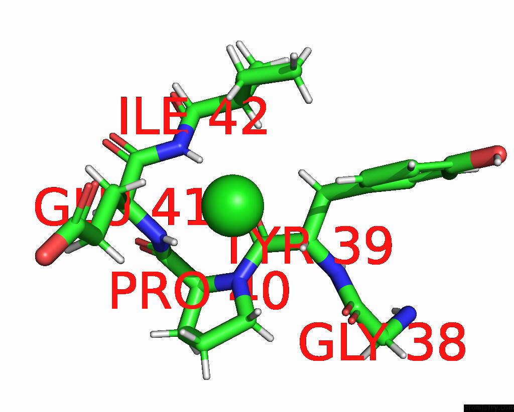
Mono view
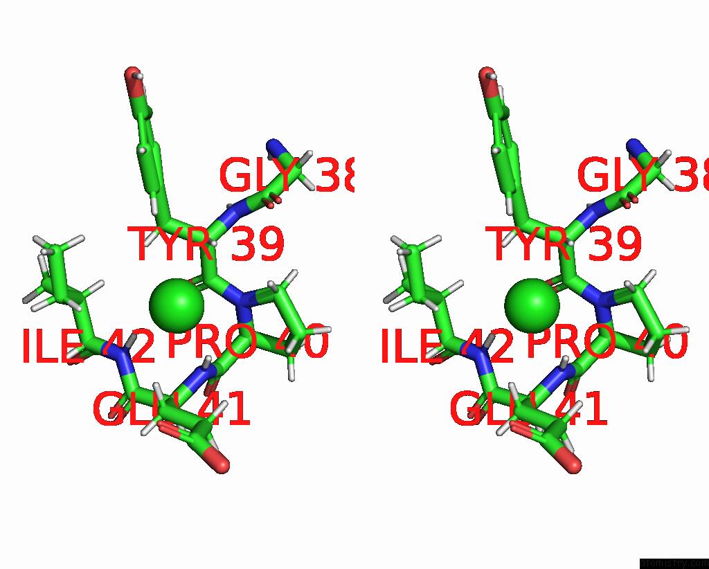
Stereo pair view
Mono view
Stereo pair view
A full contact list of Chlorine with other atoms in the Cl binding
site number 1 of Structure of Dife-Sulerythrin at 0.57 Mgy Total Absorbed Dose within 5.0Å range:
|
Chlorine binding site 2 out of 2 in 7ppu
Go back to
Chlorine binding site 2 out
of 2 in the Structure of Dife-Sulerythrin at 0.57 Mgy Total Absorbed Dose
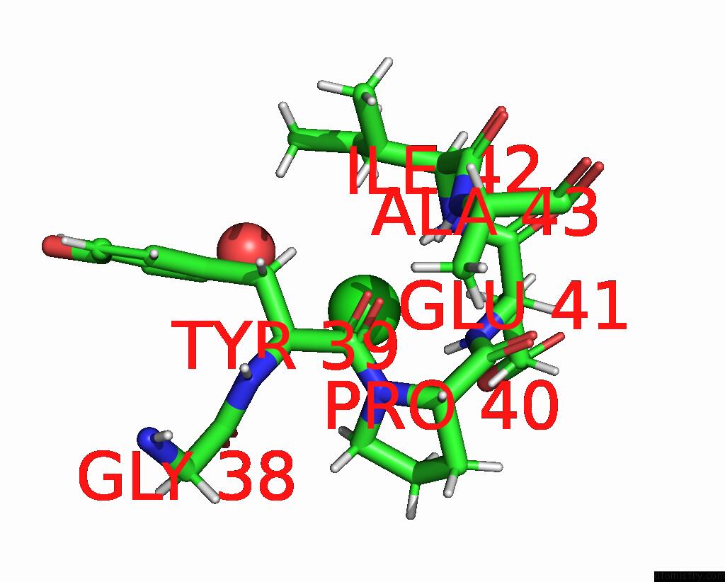
Mono view
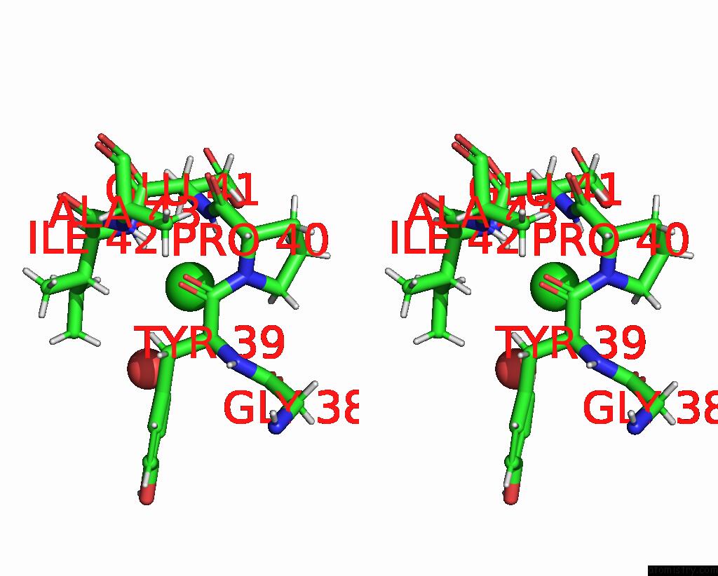
Stereo pair view
Mono view
Stereo pair view
A full contact list of Chlorine with other atoms in the Cl binding
site number 2 of Structure of Dife-Sulerythrin at 0.57 Mgy Total Absorbed Dose within 5.0Å range:
|
Reference:
F.Lennartz,
J.H.Jeoung,
S.Ruenger,
H.Dobbek,
M.S.Weiss.
Determining the Oxidation State of Elements By X-Ray Crystallography. Acta Crystallogr D Struct V. 78 238 2022BIOL.
ISSN: ISSN 2059-7983
PubMed: 35102889
DOI: 10.1107/S2059798321013048
Page generated: Sun Jul 13 05:49:44 2025
ISSN: ISSN 2059-7983
PubMed: 35102889
DOI: 10.1107/S2059798321013048
Last articles
Mn in 9LJUMn in 9LJW
Mn in 9LJS
Mn in 9LJR
Mn in 9LJT
Mn in 9LJV
Mg in 9UA2
Mg in 9R96
Mg in 9VM1
Mg in 9P01