Atomistry » Chlorine » PDB 7q1p-7q7n » 7q6m
Atomistry »
  Chlorine »
    PDB 7q1p-7q7n »
      7q6m »

Chlorine in PDB 7q6m: Structure of Wrba From Yersinia Pseudotuberculosis in P1

Enzymatic activity of Structure of Wrba From Yersinia Pseudotuberculosis in P1

All present enzymatic activity of Structure of Wrba From Yersinia Pseudotuberculosis in P1:
1.6.5.2;

Protein crystallography data

The structure of Structure of Wrba From Yersinia Pseudotuberculosis in P1, PDB code: 7q6m was solved by M.Gabrielsen, K.S.H.Beckham, A.J.Roe, with X-Ray Crystallography technique. A brief refinement statistics is given in the table below:

Resolution Low / High (Å) 43.86 / 2.04
Space group P 1
Cell size a, b, c (Å), α, β, γ (°) 60.69, 63.86, 66.36, 61.21, 77.9, 86.93
R / Rfree (%) 17.4 / 21.2

Chlorine Binding Sites:

The binding sites of Chlorine atom in the Structure of Wrba From Yersinia Pseudotuberculosis in P1 (pdb code 7q6m). This binding sites where shown within 5.0 Angstroms radius around Chlorine atom.
In total 4 binding sites of Chlorine where determined in the Structure of Wrba From Yersinia Pseudotuberculosis in P1, PDB code: 7q6m:
Jump to Chlorine binding site number: 1; 2; 3; 4;

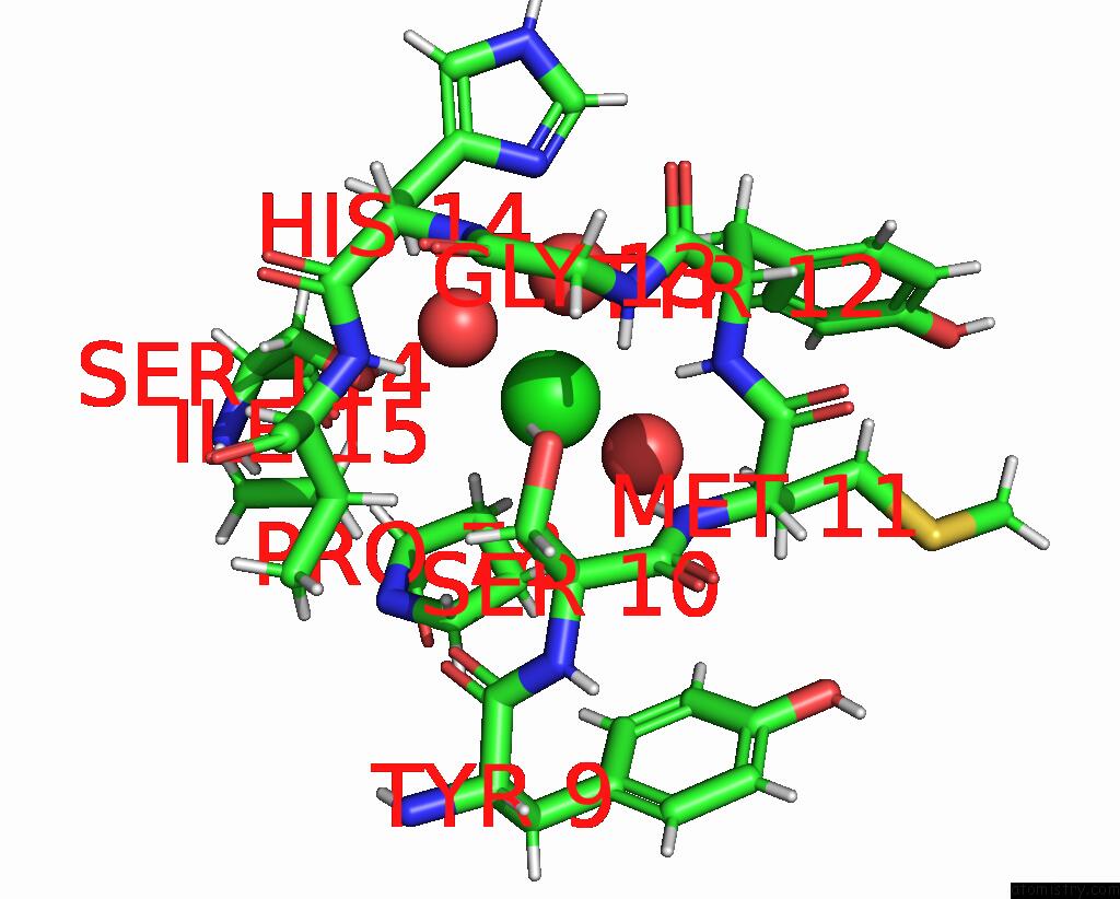
Chlorine binding site 1 out of 4 in 7q6m

Go back to Chlorine Binding Sites List in 7q6m
Chlorine binding site 1 out of 4 in the Structure of Wrba From Yersinia Pseudotuberculosis in P1


Mono view


Stereo pair view

A full contact list of Chlorine with other atoms in the Cl binding site number 1 of Structure of Wrba From Yersinia Pseudotuberculosis in P1 within 5.0Å range:
probe atom residue distance (Å) B Occ
A:Cl201

b:28.3
occ:1.00
H A:TYR12 2.6 11.3 1.0
H A:MET11 2.7 14.8 1.0
HG A:SER10 2.8 12.7 1.0
HA A:SER10 3.2 9.9 1.0
H A:ILE15 3.2 7.1 1.0
HB3 A:PRO78 3.2 6.8 1.0
O A:HOH403 3.3 19.0 1.0
N A:MET11 3.4 15.4 1.0
N A:TYR12 3.4 13.6 1.0
H A:HIS14 3.4 11.8 1.0
H A:GLY13 3.5 13.5 1.0
HG3 A:PRO78 3.5 7.9 1.0
O A:HOH374 3.6 6.7 1.0
OG A:SER10 3.6 9.3 1.0
HB2 A:HIS14 3.6 12.3 1.0
HG12 A:ILE15 3.6 9.3 1.0
HB2 A:TYR12 3.7 14.8 1.0
HB A:ILE15 3.7 7.8 1.0
HB3 A:MET11 3.8 13.2 1.0
CA A:SER10 3.9 8.2 1.0
N A:GLY13 4.0 10.4 1.0
CB A:PRO78 4.0 6.2 1.0
N A:HIS14 4.0 11.6 1.0
N A:ILE15 4.1 8.1 1.0
HD3 A:PRO78 4.1 8.4 1.0
HD2 A:TYR12 4.1 15.1 1.0
C A:SER10 4.1 10.1 1.0
CG A:PRO78 4.2 6.2 1.0
HG13 A:ILE15 4.2 9.3 1.0
ND1 A:HIS14 4.2 13.0 1.0
CG1 A:ILE15 4.2 8.3 1.0
CB A:SER10 4.2 9.4 1.0
CA A:TYR12 4.2 15.0 1.0
OG A:SER114 4.3 8.2 1.0
CA A:MET11 4.3 14.6 1.0
C A:MET11 4.3 15.5 1.0
HG A:SER114 4.3 11.6 1.0
C A:TYR12 4.4 15.2 1.0
CB A:TYR12 4.4 14.8 1.0
CB A:HIS14 4.4 11.4 1.0
HB2 A:PRO78 4.4 6.8 1.0
CB A:ILE15 4.4 8.1 1.0
HB2 A:SER10 4.4 13.4 1.0
CB A:MET11 4.5 14.9 1.0
O A:HOH313 4.6 8.0 1.0
CD A:PRO78 4.7 6.1 1.0
CA A:HIS14 4.7 11.3 1.0
CG A:HIS14 4.8 12.7 1.0
CD2 A:TYR12 4.8 14.6 1.0
C A:GLY13 4.8 10.4 1.0
C A:HIS14 4.9 10.8 1.0
CA A:ILE15 4.9 8.2 1.0
CA A:GLY13 4.9 10.8 1.0
HB2 A:MET11 5.0 13.2 1.0
O A:TYR9 5.0 7.0 1.0

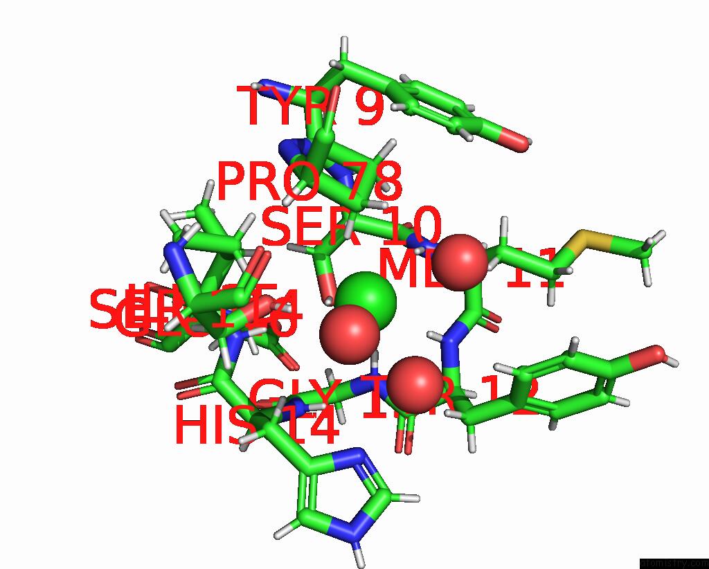
Chlorine binding site 2 out of 4 in 7q6m

Go back to Chlorine Binding Sites List in 7q6m
Chlorine binding site 2 out of 4 in the Structure of Wrba From Yersinia Pseudotuberculosis in P1


Mono view


Stereo pair view

A full contact list of Chlorine with other atoms in the Cl binding site number 2 of Structure of Wrba From Yersinia Pseudotuberculosis in P1 within 5.0Å range:
probe atom residue distance (Å) B Occ
B:Cl201

b:30.5
occ:1.00
H B:MET11 2.6 17.0 1.0
H B:TYR12 2.6 15.4 1.0
HG B:SER10 2.7 12.7 1.0
HA B:SER10 3.0 12.5 1.0
H B:ILE15 3.1 13.2 1.0
HB3 B:PRO78 3.2 9.7 1.0
N B:MET11 3.3 17.4 1.0
H B:GLY13 3.4 14.5 1.0
HG3 B:PRO78 3.4 11.2 1.0
N B:TYR12 3.5 18.2 1.0
H B:HIS14 3.5 11.1 1.0
HG12 B:ILE15 3.5 11.3 1.0
OG B:SER10 3.5 10.0 1.0
O B:HOH392 3.6 10.9 1.0
HB B:ILE15 3.6 10.7 1.0
HB2 B:HIS14 3.6 14.1 1.0
O B:HOH406 3.7 20.8 1.0
HB3 B:MET11 3.7 18.1 1.0
CA B:SER10 3.8 9.5 1.0
HB2 B:TYR12 3.9 17.5 1.0
N B:ILE15 4.0 12.1 1.0
C B:SER10 4.0 10.9 1.0
N B:GLY13 4.0 15.8 1.0
CB B:PRO78 4.0 9.2 1.0
HG13 B:ILE15 4.0 11.3 1.0
N B:HIS14 4.1 11.1 1.0
CG1 B:ILE15 4.1 11.1 1.0
CG B:PRO78 4.1 9.8 1.0
CB B:SER10 4.1 9.8 1.0
HD3 B:PRO78 4.2 11.0 1.0
HD2 B:TYR12 4.2 22.4 1.0
CA B:MET11 4.2 17.3 1.0
CB B:ILE15 4.3 11.1 1.0
OG B:SER114 4.3 11.3 1.0
ND1 B:HIS14 4.3 13.1 1.0
C B:MET11 4.3 18.3 1.0
CA B:TYR12 4.3 19.0 1.0
HB2 B:SER10 4.4 12.4 1.0
CB B:MET11 4.4 17.8 1.0
HG B:SER114 4.4 13.5 1.0
CB B:HIS14 4.4 11.7 1.0
C B:TYR12 4.5 18.7 1.0
HB2 B:PRO78 4.5 9.7 1.0
CB B:TYR12 4.6 19.0 1.0
CA B:HIS14 4.7 11.8 1.0
CD B:PRO78 4.7 9.7 1.0
O B:HOH315 4.7 8.4 1.0
HB2 B:MET11 4.8 18.1 1.0
CA B:ILE15 4.8 10.9 1.0
C B:GLY13 4.8 16.3 1.0
C B:HIS14 4.8 12.0 1.0
CD2 B:TYR12 4.9 20.3 1.0
CG B:HIS14 4.9 12.0 1.0
O B:TYR9 4.9 9.4 1.0
CA B:GLY13 4.9 17.0 1.0
HG2 B:PRO78 4.9 11.2 1.0
H B:GLU16 5.0 12.9 1.0

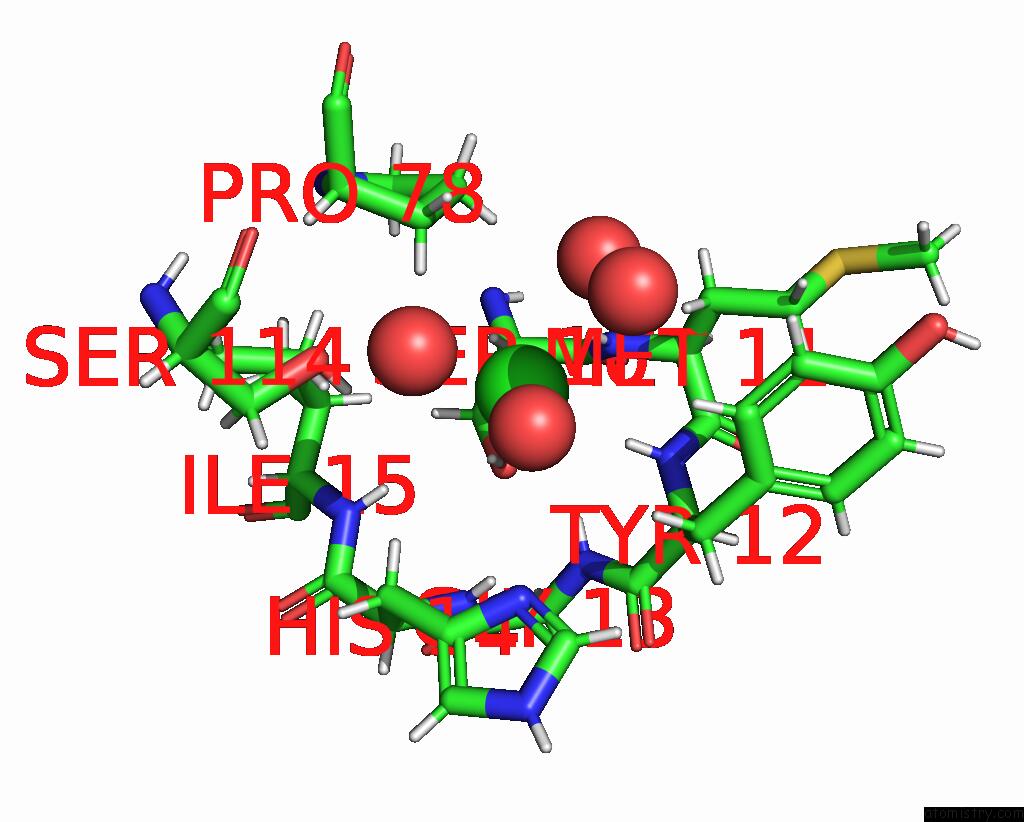
Chlorine binding site 3 out of 4 in 7q6m

Go back to Chlorine Binding Sites List in 7q6m
Chlorine binding site 3 out of 4 in the Structure of Wrba From Yersinia Pseudotuberculosis in P1


Mono view


Stereo pair view

A full contact list of Chlorine with other atoms in the Cl binding site number 3 of Structure of Wrba From Yersinia Pseudotuberculosis in P1 within 5.0Å range:
probe atom residue distance (Å) B Occ
C:Cl201

b:29.1
occ:1.00
H C:TYR12 2.5 12.7 1.0
H C:MET11 2.6 11.1 1.0
HG C:SER10 3.0 11.1 1.0
O C:HOH394 3.1 16.2 1.0
HB3 C:PRO78 3.2 8.5 1.0
HA C:SER10 3.2 8.9 1.0
O C:HOH375 3.2 6.7 1.0
N C:MET11 3.3 13.0 1.0
HG3 C:PRO78 3.3 8.2 1.0
N C:TYR12 3.3 16.0 1.0
H C:ILE15 3.5 11.0 1.0
HB3 C:MET11 3.5 12.6 1.0
H C:GLY13 3.6 13.2 1.0
HB2 C:TYR12 3.6 15.5 1.0
HG C:SER114 3.7 12.0 1.0
H C:HIS14 3.7 10.5 1.0
OG C:SER10 3.8 7.9 1.0
HG12 C:ILE15 3.8 7.6 1.0
HD2 C:TYR12 3.8 16.0 1.0
HB2 C:HIS14 3.9 11.3 1.0
CB C:PRO78 3.9 6.5 1.0
CA C:SER10 4.0 7.2 1.0
HB C:ILE15 4.0 9.1 1.0
CG C:PRO78 4.0 6.6 1.0
C C:SER10 4.1 8.3 1.0
CA C:MET11 4.1 13.2 1.0
N C:GLY13 4.1 10.9 1.0
CA C:TYR12 4.2 16.8 1.0
C C:MET11 4.2 14.7 1.0
HD3 C:PRO78 4.2 7.0 1.0
CB C:MET11 4.3 13.5 1.0
HB2 C:PRO78 4.3 8.5 1.0
CB C:TYR12 4.3 17.0 1.0
ND1 C:HIS14 4.3 11.8 1.0
N C:HIS14 4.3 10.2 1.0
HG13 C:ILE15 4.3 7.6 1.0
N C:ILE15 4.3 10.0 1.0
OG C:SER114 4.4 8.6 1.0
CB C:SER10 4.4 7.5 1.0
CG1 C:ILE15 4.4 8.4 1.0
C C:TYR12 4.4 17.7 1.0
O C:HOH310 4.5 7.8 1.0
CD2 C:TYR12 4.5 16.9 1.0
CB C:ILE15 4.6 9.2 1.0
HB2 C:SER10 4.6 9.1 1.0
CB C:HIS14 4.7 10.1 1.0
HB2 C:MET11 4.7 12.6 1.0
CD C:PRO78 4.7 6.2 1.0
O C:HOH408 4.8 23.8 1.0
HG2 C:PRO78 4.8 8.2 1.0
CG C:TYR12 4.8 17.0 1.0
CA C:HIS14 4.9 9.6 1.0
CG C:HIS14 5.0 11.0 1.0
HA C:MET11 5.0 11.1 1.0

Chlorine binding site 4 out of 4 in 7q6m

Go back to Chlorine Binding Sites List in 7q6m
Chlorine binding site 4 out of 4 in the Structure of Wrba From Yersinia Pseudotuberculosis in P1


Mono view


Stereo pair view

A full contact list of Chlorine with other atoms in the Cl binding site number 4 of Structure of Wrba From Yersinia Pseudotuberculosis in P1 within 5.0Å range:
probe atom residue distance (Å) B Occ
D:Cl201

b:33.0
occ:1.00
H D:TYR12 2.5 19.9 1.0
H D:MET11 2.6 16.5 1.0
HG D:SER10 2.8 12.7 1.0
HA D:SER10 3.2 11.6 1.0
H D:ILE15 3.2 11.8 1.0
HB3 D:PRO78 3.3 10.0 1.0
N D:MET11 3.3 16.7 1.0
N D:TYR12 3.4 20.2 1.0
H D:GLY13 3.4 15.6 1.0
HG3 D:PRO78 3.4 10.1 1.0
H D:HIS14 3.5 13.3 1.0
O D:HOH413 3.5 22.1 1.0
O D:HOH374 3.5 11.9 1.0
OG D:SER10 3.5 11.1 1.0
HB2 D:HIS14 3.6 16.0 1.0
HG12 D:ILE15 3.7 10.7 1.0
HB3 D:MET11 3.7 16.5 1.0
HB2 D:TYR12 3.8 19.1 1.0
HB D:ILE15 3.8 11.9 1.0
CA D:SER10 3.9 10.7 1.0
HD2 D:TYR12 3.9 22.6 1.0
N D:GLY13 4.0 14.1 1.0
CB D:PRO78 4.0 9.0 1.0
C D:SER10 4.1 12.5 1.0
N D:HIS14 4.1 12.9 1.0
N D:ILE15 4.1 10.6 1.0
CG D:PRO78 4.1 9.2 1.0
CA D:MET11 4.2 16.3 1.0
HG13 D:ILE15 4.2 10.7 1.0
CA D:TYR12 4.2 19.4 1.0
CB D:SER10 4.3 11.5 1.0
C D:MET11 4.3 18.2 1.0
CG1 D:ILE15 4.3 9.9 1.0
HD3 D:PRO78 4.3 9.6 1.0
ND1 D:HIS14 4.3 14.4 1.0
OG D:SER114 4.4 9.4 1.0
CB D:MET11 4.4 16.8 1.0
C D:TYR12 4.4 20.4 1.0
CB D:TYR12 4.4 20.0 1.0
HG D:SER114 4.4 13.3 1.0
CB D:HIS14 4.4 13.3 1.0
CB D:ILE15 4.4 10.5 1.0
HB2 D:PRO78 4.5 10.0 1.0
HB2 D:SER10 4.5 13.9 1.0
O D:HOH311 4.6 9.9 1.0
CD2 D:TYR12 4.7 20.9 1.0
CA D:HIS14 4.7 12.9 1.0
CD D:PRO78 4.7 9.0 1.0
HB2 D:MET11 4.8 16.5 1.0
C D:GLY13 4.8 13.9 1.0
CG D:HIS14 4.9 14.5 1.0
CA D:GLY13 4.9 14.3 1.0
C D:HIS14 4.9 13.0 1.0
HG2 D:PRO78 4.9 10.1 1.0
CA D:ILE15 4.9 10.6 1.0
CG D:TYR12 5.0 20.0 1.0

Reference:

R.Zambelloni, K.S.H.Beckham, H.J.Wu, M.Elofsson, R.Marquez, M.Gabrielsen, A.J.Roe. Crystal Structures of Wrba, A Spurious Target of the Salicylidene Acylhydrazide Inhibitors of Type III Secretion in Gram-Negative Pathogens, and Verification of Improved Specificity of Next-Generation Compounds. Microbiology (Reading, V. 168 2022ENGL.).
ISSN: ESSN 1465-2080
PubMed: 35829699
DOI: 10.1099/MIC.0.001211
Page generated: Sun Jul 13 06:11:32 2025

Last articles

Na in 3CCE
Na in 3CC7
Na in 3CC4
Na in 3CC9
Na in 3CBC
Na in 3CBT
Na in 3C9F
Na in 3CB8
Na in 3C7O
Na in 3C88
© Copyright 2008-2020 by atomistry.com
Home   |    Site Map   |    Copyright   |    Contact us   |    Privacy