Chlorine »
PDB 7t4t-7thh »
7t88 »
Chlorine in PDB 7t88: Crystal Structure of the C-Terminal Domain of the Phosphate Acetyltransferase From Escherichia Coli
Enzymatic activity of Crystal Structure of the C-Terminal Domain of the Phosphate Acetyltransferase From Escherichia Coli
All present enzymatic activity of Crystal Structure of the C-Terminal Domain of the Phosphate Acetyltransferase From Escherichia Coli:
2.3.1.8;
2.3.1.8;
Protein crystallography data
The structure of Crystal Structure of the C-Terminal Domain of the Phosphate Acetyltransferase From Escherichia Coli, PDB code: 7t88
was solved by
Y.Kim,
A.Dementiev,
L.Welk,
M.Endres,
A.Joachimiak,
Center For Structuralgenomics Of Infectious Diseases (Csgid),
with X-Ray Crystallography technique. A brief refinement statistics is given in the table below:
| Resolution Low / High (Å) | 48.26 / 2.10 |
| Space group | P 41 21 2 |
| Cell size a, b, c (Å), α, β, γ (°) | 72.875, 72.875, 193.248, 90, 90, 90 |
| R / Rfree (%) | 20 / 23.1 |
Other elements in 7t88:
The structure of Crystal Structure of the C-Terminal Domain of the Phosphate Acetyltransferase From Escherichia Coli also contains other interesting chemical elements:
| Sodium | (Na) | 1 atom |
| Potassium | (K) | 1 atom |
| Iodine | (I) | 2 atoms |
Chlorine Binding Sites:
The binding sites of Chlorine atom in the Crystal Structure of the C-Terminal Domain of the Phosphate Acetyltransferase From Escherichia Coli
(pdb code 7t88). This binding sites where shown within
5.0 Angstroms radius around Chlorine atom.
In total only one binding site of Chlorine was determined in the Crystal Structure of the C-Terminal Domain of the Phosphate Acetyltransferase From Escherichia Coli, PDB code: 7t88:
In total only one binding site of Chlorine was determined in the Crystal Structure of the C-Terminal Domain of the Phosphate Acetyltransferase From Escherichia Coli, PDB code: 7t88:
Chlorine binding site 1 out of 1 in 7t88
Go back to
Chlorine binding site 1 out
of 1 in the Crystal Structure of the C-Terminal Domain of the Phosphate Acetyltransferase From Escherichia Coli
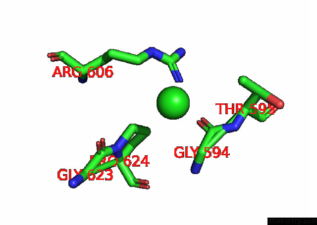
Mono view
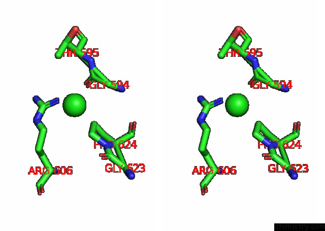
Stereo pair view
Mono view
Stereo pair view
A full contact list of Chlorine with other atoms in the Cl binding
site number 1 of Crystal Structure of the C-Terminal Domain of the Phosphate Acetyltransferase From Escherichia Coli within 5.0Å range:
|
Reference:
Y.Kim,
A.Dementiev,
L.Welk,
M.Endres,
A.Joachimiak,
Center For Structural Genomics Of Infectious Diseases(Csgid).
Crystal Structure of C From Escherichia Coli To Be Published.
Page generated: Sun Jul 13 07:22:27 2025
Last articles
Na in 7AC2Na in 7A76
Na in 7AAW
Na in 7A6Q
Na in 7A74
Na in 7A70
Na in 7A56
Na in 7A3K
Na in 7A68
Na in 7A3I