Chlorine »
PDB 7u24-7uer »
7u56 »
Chlorine in PDB 7u56: Crystal Structure of D-Alanine--D-Alanine Ligase From Klebsiella Pneumoniae Subsp. Pneumoniae in Complex with Amp
Enzymatic activity of Crystal Structure of D-Alanine--D-Alanine Ligase From Klebsiella Pneumoniae Subsp. Pneumoniae in Complex with Amp
All present enzymatic activity of Crystal Structure of D-Alanine--D-Alanine Ligase From Klebsiella Pneumoniae Subsp. Pneumoniae in Complex with Amp:
6.3.2.4;
6.3.2.4;
Protein crystallography data
The structure of Crystal Structure of D-Alanine--D-Alanine Ligase From Klebsiella Pneumoniae Subsp. Pneumoniae in Complex with Amp, PDB code: 7u56
was solved by
Seattle Structural Genomics Center For Infectious Disease (Ssgcid),
with X-Ray Crystallography technique. A brief refinement statistics is given in the table below:
| Resolution Low / High (Å) | 39.13 / 1.85 |
| Space group | P 21 21 2 |
| Cell size a, b, c (Å), α, β, γ (°) | 95.35, 115.42, 68.48, 90, 90, 90 |
| R / Rfree (%) | 16.2 / 19.9 |
Other elements in 7u56:
The structure of Crystal Structure of D-Alanine--D-Alanine Ligase From Klebsiella Pneumoniae Subsp. Pneumoniae in Complex with Amp also contains other interesting chemical elements:
| Magnesium | (Mg) | 2 atoms |
Chlorine Binding Sites:
The binding sites of Chlorine atom in the Crystal Structure of D-Alanine--D-Alanine Ligase From Klebsiella Pneumoniae Subsp. Pneumoniae in Complex with Amp
(pdb code 7u56). This binding sites where shown within
5.0 Angstroms radius around Chlorine atom.
In total 4 binding sites of Chlorine where determined in the Crystal Structure of D-Alanine--D-Alanine Ligase From Klebsiella Pneumoniae Subsp. Pneumoniae in Complex with Amp, PDB code: 7u56:
Jump to Chlorine binding site number: 1; 2; 3; 4;
In total 4 binding sites of Chlorine where determined in the Crystal Structure of D-Alanine--D-Alanine Ligase From Klebsiella Pneumoniae Subsp. Pneumoniae in Complex with Amp, PDB code: 7u56:
Jump to Chlorine binding site number: 1; 2; 3; 4;
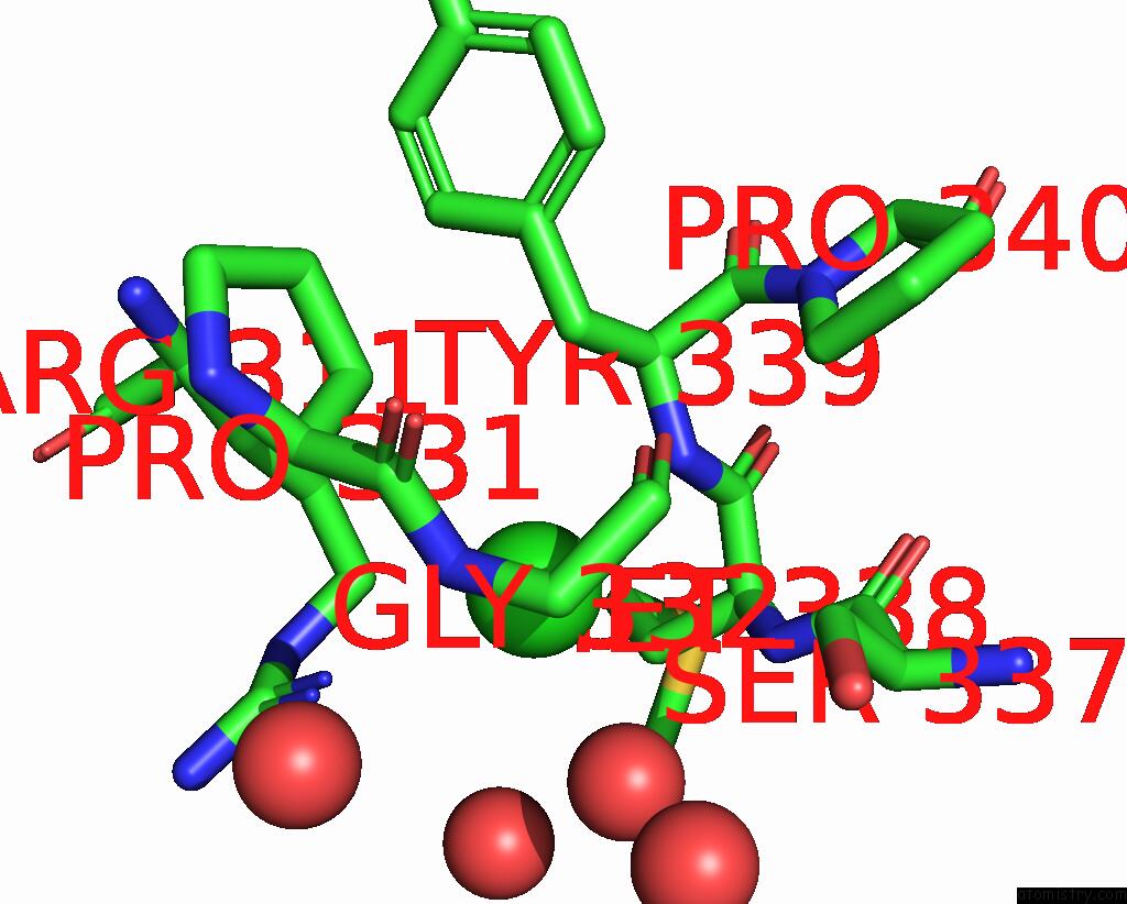
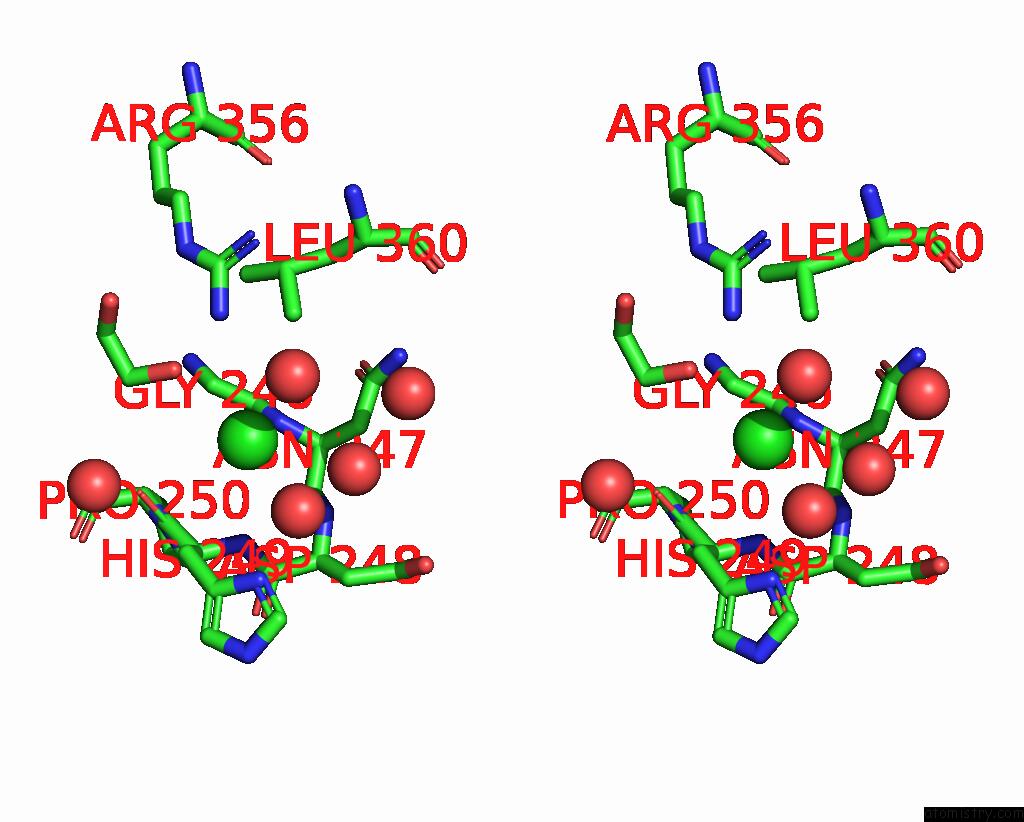
Chlorine binding site 1 out of 4 in 7u56
Go back to
Chlorine binding site 1 out
of 4 in the Crystal Structure of D-Alanine--D-Alanine Ligase From Klebsiella Pneumoniae Subsp. Pneumoniae in Complex with Amp
Mono view

Stereo pair view
Mono view
Stereo pair view
A full contact list of Chlorine with other atoms in the Cl binding
site number 1 of Crystal Structure of D-Alanine--D-Alanine Ligase From Klebsiella Pneumoniae Subsp. Pneumoniae in Complex with Amp within 5.0Å range:
|
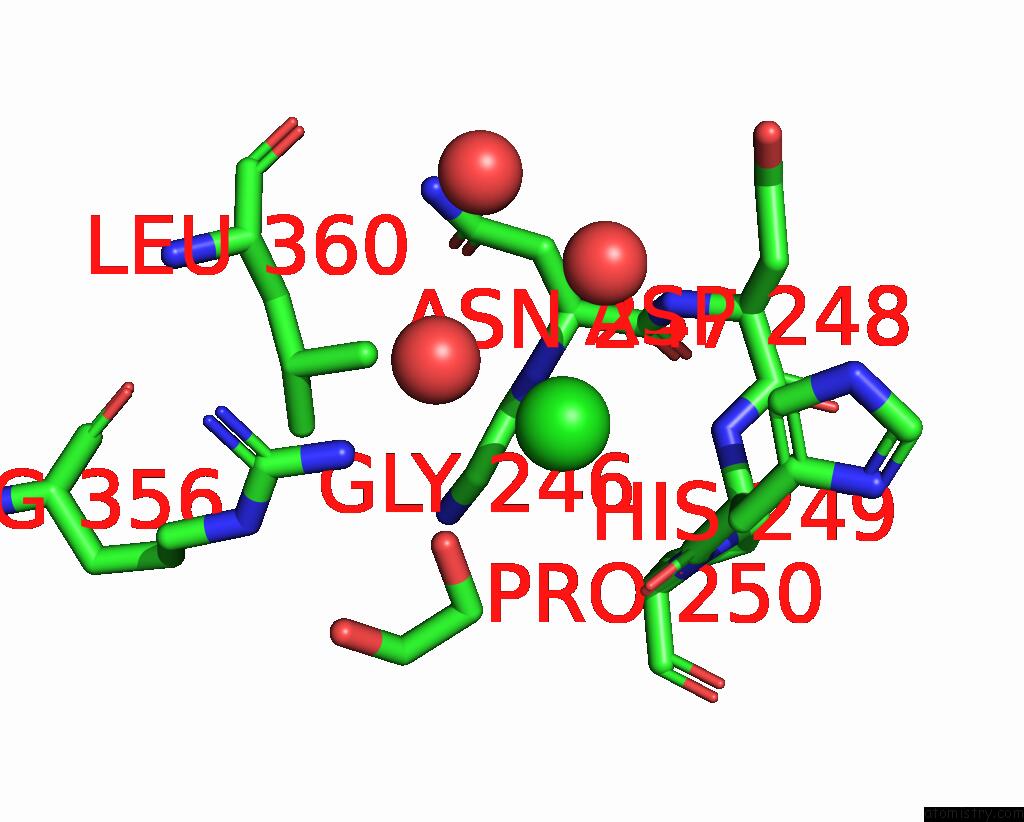
Chlorine binding site 2 out of 4 in 7u56
Go back to
Chlorine binding site 2 out
of 4 in the Crystal Structure of D-Alanine--D-Alanine Ligase From Klebsiella Pneumoniae Subsp. Pneumoniae in Complex with Amp
Mono view

Stereo pair view
Mono view
Stereo pair view
A full contact list of Chlorine with other atoms in the Cl binding
site number 2 of Crystal Structure of D-Alanine--D-Alanine Ligase From Klebsiella Pneumoniae Subsp. Pneumoniae in Complex with Amp within 5.0Å range:
|
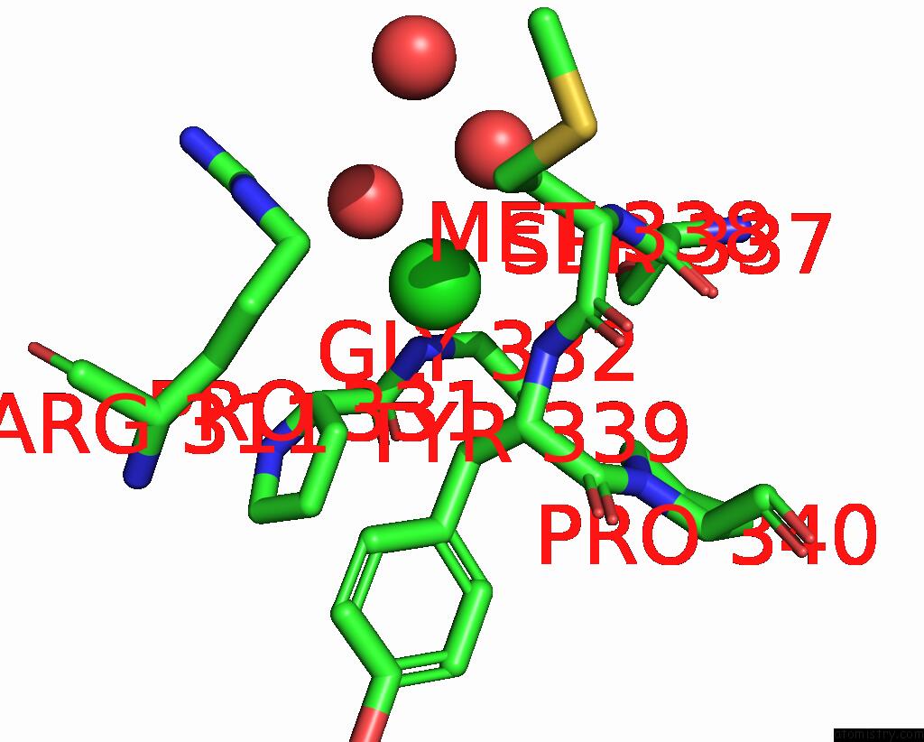
Chlorine binding site 3 out of 4 in 7u56
Go back to
Chlorine binding site 3 out
of 4 in the Crystal Structure of D-Alanine--D-Alanine Ligase From Klebsiella Pneumoniae Subsp. Pneumoniae in Complex with Amp
Mono view

Stereo pair view
Mono view
Stereo pair view
A full contact list of Chlorine with other atoms in the Cl binding
site number 3 of Crystal Structure of D-Alanine--D-Alanine Ligase From Klebsiella Pneumoniae Subsp. Pneumoniae in Complex with Amp within 5.0Å range:
|
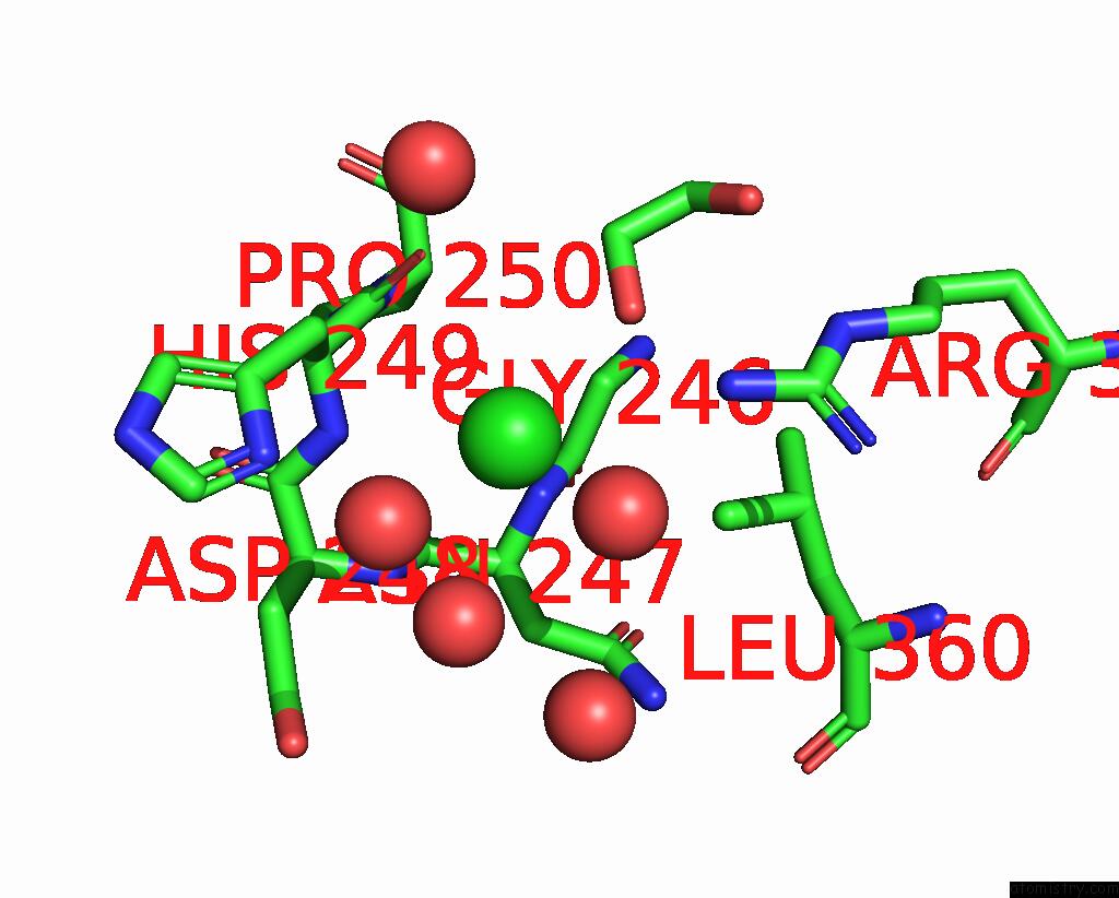
Chlorine binding site 4 out of 4 in 7u56
Go back to
Chlorine binding site 4 out
of 4 in the Crystal Structure of D-Alanine--D-Alanine Ligase From Klebsiella Pneumoniae Subsp. Pneumoniae in Complex with Amp
Mono view
Stereo pair view
Mono view
Stereo pair view
A full contact list of Chlorine with other atoms in the Cl binding
site number 4 of Crystal Structure of D-Alanine--D-Alanine Ligase From Klebsiella Pneumoniae Subsp. Pneumoniae in Complex with Amp within 5.0Å range:
|
Reference:
J.Abendroth,
M.J.Bolejack,
D.D.Lorimer,
P.S.Horanyi,
T.E.Edwards.
Crystal Structure of D-Alanine--D-Alanine Ligase From Klebsiella Pneumoniae Subsp. Pneumoniae in Complex with Amp To Be Published.
Page generated: Sun Jul 13 07:43:28 2025
Last articles
Mg in 5S53Mg in 5S54
Mg in 5S55
Mg in 5S52
Mg in 5S50
Mg in 5S51
Mg in 5S4Y
Mg in 5S4Z
Mg in 5S4X
Mg in 5S4V