Chlorine »
PDB 8b7u-8bid »
8beo »
Chlorine in PDB 8beo: Crystal Structure of E. Coli Glyoxylate Carboligase Mutant I393A with Map
Enzymatic activity of Crystal Structure of E. Coli Glyoxylate Carboligase Mutant I393A with Map
All present enzymatic activity of Crystal Structure of E. Coli Glyoxylate Carboligase Mutant I393A with Map:
4.1.1.47;
4.1.1.47;
Protein crystallography data
The structure of Crystal Structure of E. Coli Glyoxylate Carboligase Mutant I393A with Map, PDB code: 8beo
was solved by
B.Shaanan,
E.Binshtein,
with X-Ray Crystallography technique. A brief refinement statistics is given in the table below:
| Resolution Low / High (Å) | 48.35 / 1.96 |
| Space group | P 41 21 2 |
| Cell size a, b, c (Å), α, β, γ (°) | 188.641, 188.641, 246.957, 90, 90, 90 |
| R / Rfree (%) | 17.6 / 20.4 |
Other elements in 8beo:
The structure of Crystal Structure of E. Coli Glyoxylate Carboligase Mutant I393A with Map also contains other interesting chemical elements:
| Sodium | (Na) | 15 atoms |
| Magnesium | (Mg) | 13 atoms |
Chlorine Binding Sites:
The binding sites of Chlorine atom in the Crystal Structure of E. Coli Glyoxylate Carboligase Mutant I393A with Map
(pdb code 8beo). This binding sites where shown within
5.0 Angstroms radius around Chlorine atom.
In total only one binding site of Chlorine was determined in the Crystal Structure of E. Coli Glyoxylate Carboligase Mutant I393A with Map, PDB code: 8beo:
In total only one binding site of Chlorine was determined in the Crystal Structure of E. Coli Glyoxylate Carboligase Mutant I393A with Map, PDB code: 8beo:
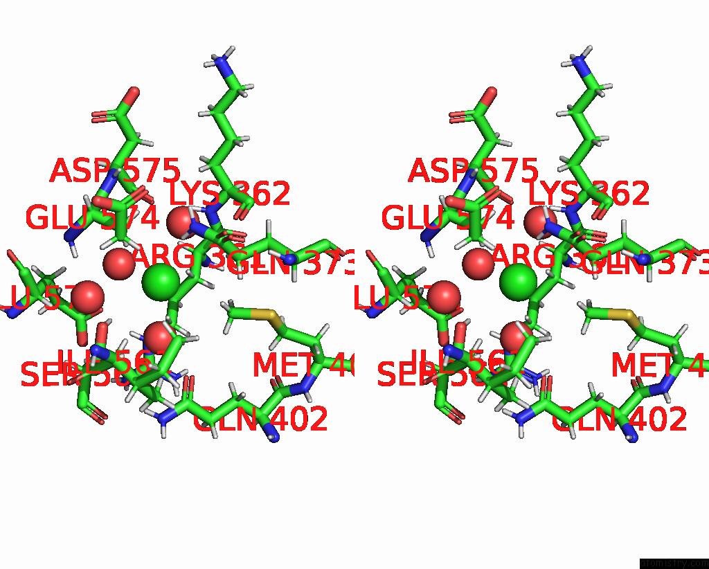
Chlorine binding site 1 out of 1 in 8beo
Go back to
Chlorine binding site 1 out
of 1 in the Crystal Structure of E. Coli Glyoxylate Carboligase Mutant I393A with Map

Mono view
Stereo pair view
Mono view
Stereo pair view
A full contact list of Chlorine with other atoms in the Cl binding
site number 1 of Crystal Structure of E. Coli Glyoxylate Carboligase Mutant I393A with Map within 5.0Å range:
|
Reference:
B.Shaanan,
E.Binshtein.
Crystal Structure of E. Coli Glyoxylate Carboligase Mutant I393A with Map To Be Published.
Page generated: Sun Jul 13 09:34:50 2025
Last articles
Mg in 2R3BMg in 2R5T
Mg in 2R42
Mg in 2R20
Mg in 2R25
Mg in 2R2H
Mg in 2R23
Mg in 2R1X
Mg in 2R1W
Mg in 2R1Y