Chlorine »
PDB 8c4j-8ccb »
8c5d »
Chlorine in PDB 8c5d: Glutathione Transferase P1-1 From Mus Musculus
Enzymatic activity of Glutathione Transferase P1-1 From Mus Musculus
All present enzymatic activity of Glutathione Transferase P1-1 From Mus Musculus:
2.5.1.18;
2.5.1.18;
Protein crystallography data
The structure of Glutathione Transferase P1-1 From Mus Musculus, PDB code: 8c5d
was solved by
A.C.Papageorgiou,
with X-Ray Crystallography technique. A brief refinement statistics is given in the table below:
| Resolution Low / High (Å) | 38.68 / 1.28 |
| Space group | P 21 21 21 |
| Cell size a, b, c (Å), α, β, γ (°) | 56.617, 77.366, 101.444, 90, 90, 90 |
| R / Rfree (%) | 17.5 / 20 |
Other elements in 8c5d:
The structure of Glutathione Transferase P1-1 From Mus Musculus also contains other interesting chemical elements:
| Sodium | (Na) | 11 atoms |
| Calcium | (Ca) | 4 atoms |
Chlorine Binding Sites:
The binding sites of Chlorine atom in the Glutathione Transferase P1-1 From Mus Musculus
(pdb code 8c5d). This binding sites where shown within
5.0 Angstroms radius around Chlorine atom.
In total only one binding site of Chlorine was determined in the Glutathione Transferase P1-1 From Mus Musculus, PDB code: 8c5d:
In total only one binding site of Chlorine was determined in the Glutathione Transferase P1-1 From Mus Musculus, PDB code: 8c5d:
Chlorine binding site 1 out of 1 in 8c5d
Go back to
Chlorine binding site 1 out
of 1 in the Glutathione Transferase P1-1 From Mus Musculus

Mono view
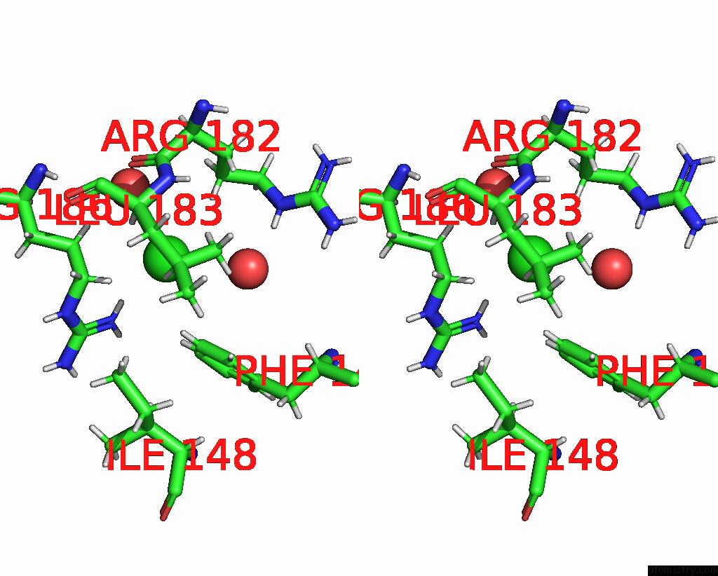
Stereo pair view
Mono view
Stereo pair view
A full contact list of Chlorine with other atoms in the Cl binding
site number 1 of Glutathione Transferase P1-1 From Mus Musculus within 5.0Å range:
|
Reference:
O.Kupreienko,
F.Pouliou,
K.Konstandinidis,
I.Axarli,
E.Douni,
A.C.Papageorgiou,
N.E.Labrou.
Inhibition Analysis and High-Resolution Crystal Structure of Mus Musculus Glutathione Transferase P1-1. Biomolecules V. 13 2023.
ISSN: ESSN 2218-273X
PubMed: 37189361
DOI: 10.3390/BIOM13040613
Page generated: Sun Jul 13 09:59:43 2025
ISSN: ESSN 2218-273X
PubMed: 37189361
DOI: 10.3390/BIOM13040613
Last articles
Pt in 5T7LPt in 5NJ7
Pt in 5OLD
Pt in 5OLE
Pt in 5N26
Pt in 5MU1
Pt in 5NA9
Pt in 5NJ1
Pt in 5L4R
Pt in 5MIK