Atomistry » Chlorine » PDB 8cvu-8d1l » 8cwd
Atomistry »
  Chlorine »
    PDB 8cvu-8d1l »
      8cwd »

Chlorine in PDB 8cwd: 20NS Temperature-Jump (DARK1) Xfel Structure of Lysozyme Bound to N, N'-Diacetylchitobiose

Enzymatic activity of 20NS Temperature-Jump (DARK1) Xfel Structure of Lysozyme Bound to N, N'-Diacetylchitobiose

All present enzymatic activity of 20NS Temperature-Jump (DARK1) Xfel Structure of Lysozyme Bound to N, N'-Diacetylchitobiose:
3.2.1.17;

Protein crystallography data

The structure of 20NS Temperature-Jump (DARK1) Xfel Structure of Lysozyme Bound to N, N'-Diacetylchitobiose, PDB code: 8cwd was solved by A.M.Wolff, M.C.Thompson, J.S.Fraser, E.Nango, with X-Ray Crystallography technique. A brief refinement statistics is given in the table below:

Resolution Low / High (Å) 30.74 / 1.48
Space group P 43 21 2
Cell size a, b, c (Å), α, β, γ (°) 77.061, 77.061, 37.223, 90, 90, 90
R / Rfree (%) 13 / 16.3

Other elements in 8cwd:

The structure of 20NS Temperature-Jump (DARK1) Xfel Structure of Lysozyme Bound to N, N'-Diacetylchitobiose also contains other interesting chemical elements:

Sodium (Na) 2 atoms

Chlorine Binding Sites:

The binding sites of Chlorine atom in the 20NS Temperature-Jump (DARK1) Xfel Structure of Lysozyme Bound to N, N'-Diacetylchitobiose (pdb code 8cwd). This binding sites where shown within 5.0 Angstroms radius around Chlorine atom.
In total 5 binding sites of Chlorine where determined in the 20NS Temperature-Jump (DARK1) Xfel Structure of Lysozyme Bound to N, N'-Diacetylchitobiose, PDB code: 8cwd:
Jump to Chlorine binding site number: 1; 2; 3; 4; 5;

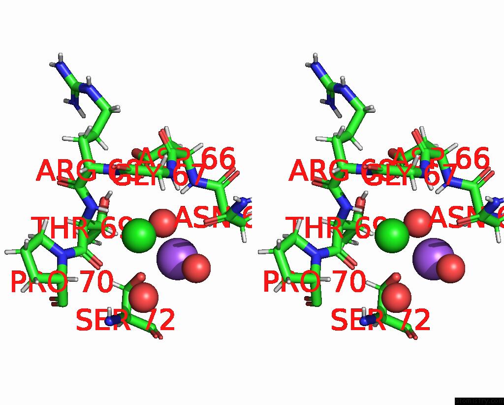
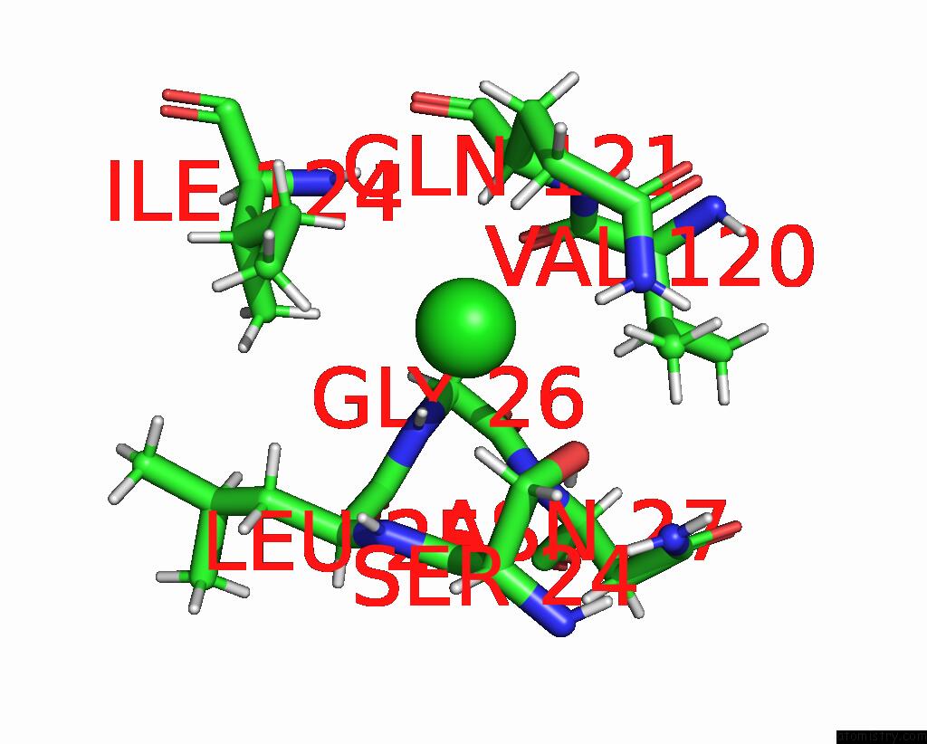
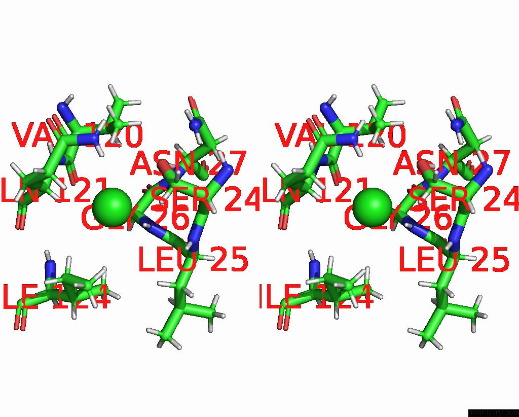
Chlorine binding site 1 out of 5 in 8cwd

Go back to Chlorine Binding Sites List in 8cwd
Chlorine binding site 1 out of 5 in the 20NS Temperature-Jump (DARK1) Xfel Structure of Lysozyme Bound to N, N'-Diacetylchitobiose


Mono view


Stereo pair view

A full contact list of Chlorine with other atoms in the Cl binding site number 1 of 20NS Temperature-Jump (DARK1) Xfel Structure of Lysozyme Bound to N, N'-Diacetylchitobiose within 5.0Å range:
probe atom residue distance (Å) B Occ
A:Cl204

b:32.2
occ:1.00
H A:THR69 2.4 19.6 1.0
O A:HOH350 3.0 18.1 1.0
H A:GLY67 3.2 20.0 1.0
O A:THR69 3.2 18.8 1.0
O A:HOH384 3.2 36.3 1.0
N A:THR69 3.2 16.3 1.0
HA2 A:GLY67 3.2 23.2 1.0
H A:ARG68 3.4 19.7 1.0
C A:THR69 3.5 17.0 1.0
OD1 A:ASN65 3.5 25.0 1.0
N A:ARG68 3.5 16.4 1.0
N A:GLY67 3.6 16.7 1.0
HB A:THR69 3.6 17.8 1.0
C A:GLY67 3.6 18.5 1.0
CA A:GLY67 3.6 19.3 1.0
OG A:SER72 3.7 18.9 1.0
CA A:THR69 3.8 16.4 1.0
O A:HOH380 3.9 20.5 1.0
HA A:PRO70 3.9 25.3 1.0
H A:ASP66 4.0 17.1 1.0
OD1 A:ASP66 4.1 13.9 1.0
N A:PRO70 4.1 18.2 1.0
CB A:THR69 4.2 14.9 1.0
C A:ARG68 4.2 18.5 1.0
O A:GLY67 4.3 22.6 1.0
CA A:ARG68 4.3 17.4 1.0
HG1 A:THR69 4.4 18.1 1.0
HA A:ARG68 4.5 20.8 1.0
CA A:PRO70 4.5 21.1 1.0
HA3 A:GLY67 4.6 23.2 1.0
HA A:ASN65 4.6 17.6 1.0
H A:SER72 4.6 25.2 1.0
C A:ASP66 4.6 15.9 1.0
N A:ASP66 4.7 14.3 1.0
NA A:NA201 4.7 16.6 1.0
CG A:ASN65 4.7 23.0 1.0
HA A:THR69 4.7 19.7 1.0
OG1 A:THR69 4.8 15.1 1.0
HD3 A:PRO70 4.8 26.1 1.0
CB A:SER72 5.0 18.7 1.0

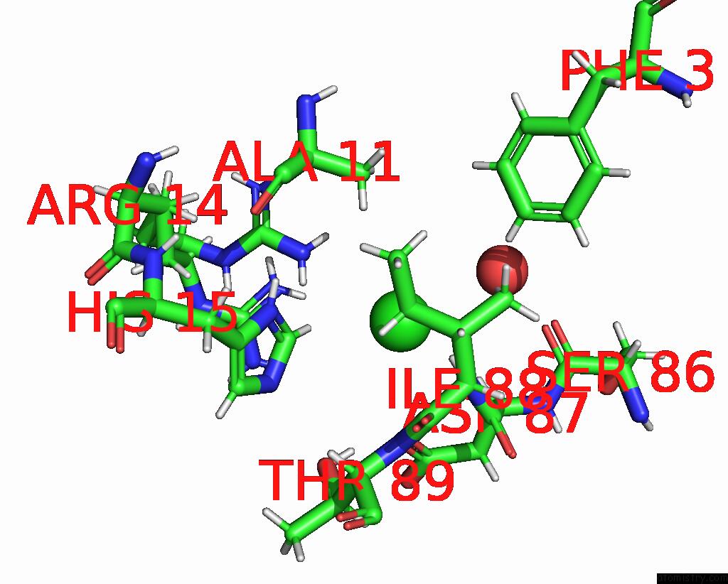
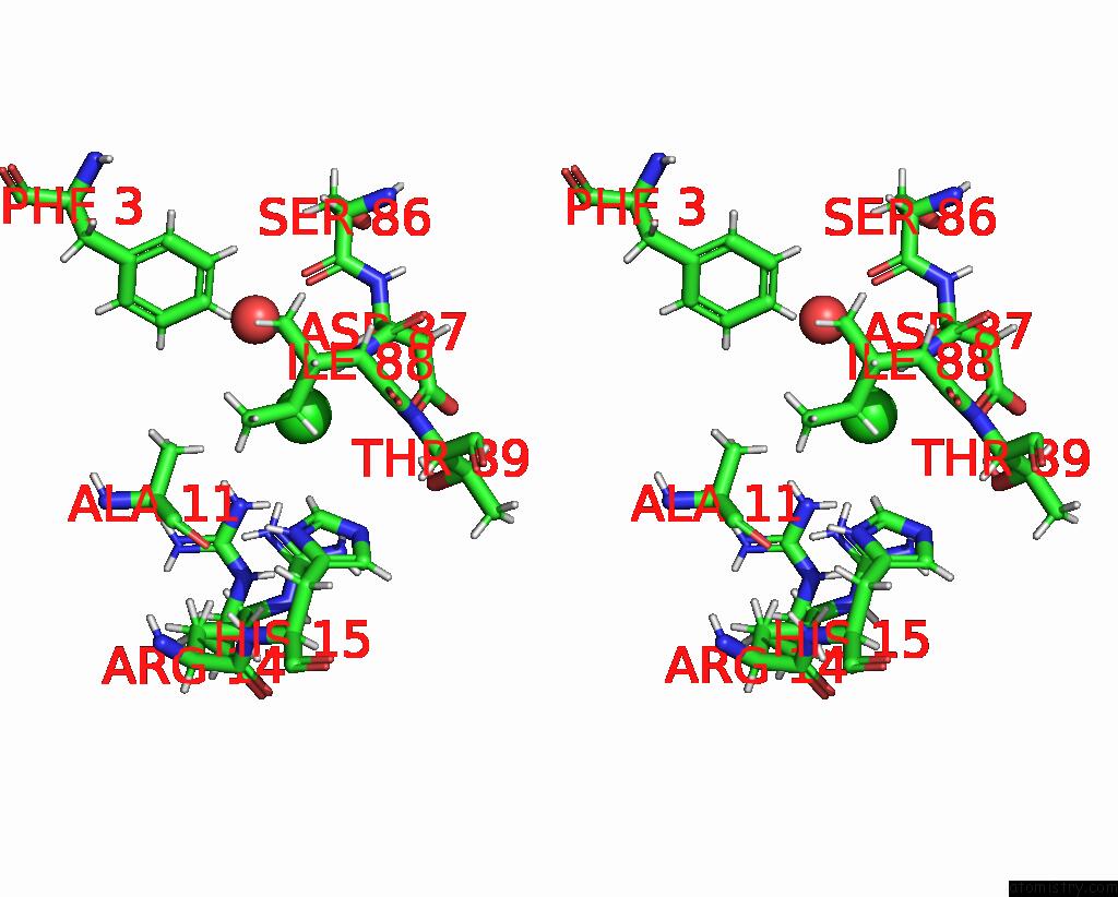
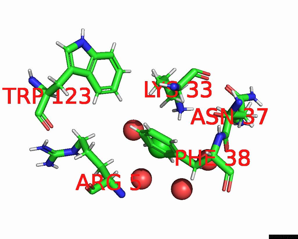
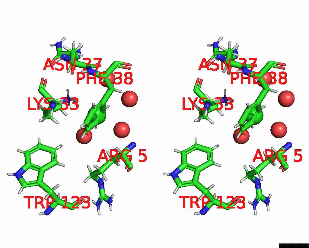
Chlorine binding site 2 out of 5 in 8cwd

Go back to Chlorine Binding Sites List in 8cwd
Chlorine binding site 2 out of 5 in the 20NS Temperature-Jump (DARK1) Xfel Structure of Lysozyme Bound to N, N'-Diacetylchitobiose


Mono view


Stereo pair view

A full contact list of Chlorine with other atoms in the Cl binding site number 2 of 20NS Temperature-Jump (DARK1) Xfel Structure of Lysozyme Bound to N, N'-Diacetylchitobiose within 5.0Å range:
probe atom residue distance (Å) B Occ
A:Cl205

b:87.1
occ:1.00
H A:ILE88 2.3 21.3 1.0
HG12 A:ILE88 2.6 21.6 1.0
HE1 A:HIS15 2.7 35.1 1.0
HA A:ASP87 2.9 25.2 1.0
N A:ILE88 3.1 17.8 1.0
HH21 A:ARG14 3.2 33.4 0.4
HH22 A:ARG14 3.4 33.4 0.4
OD1 A:ASP87 3.5 28.3 1.0
CE1 A:HIS15 3.5 29.3 1.0
CG1 A:ILE88 3.6 18.0 1.0
NH2 A:ARG14 3.6 27.9 0.4
CA A:ASP87 3.7 21.0 1.0
HH12 A:ARG14 3.7 40.4 0.6
HG23 A:ILE88 3.8 22.6 1.0
HG13 A:ILE88 3.8 21.6 1.0
HB1 A:ALA11 3.8 21.9 1.0
C A:ASP87 3.9 18.4 1.0
HZ A:PHE3 3.9 21.8 1.0
O A:HOH339 3.9 34.3 1.0
H A:THR89 4.0 19.4 1.0
HG1 A:THR89 4.0 26.9 1.0
CA A:ILE88 4.1 15.6 1.0
CG A:ASP87 4.1 29.9 1.0
CB A:ILE88 4.2 16.1 1.0
O A:SER86 4.3 24.4 1.0
NE2 A:HIS15 4.3 31.1 1.0
ND1 A:HIS15 4.3 28.8 1.0
NH1 A:ARG14 4.4 33.7 0.6
HD1 A:HIS15 4.4 34.5 1.0
HB2 A:ALA11 4.4 21.9 1.0
N A:THR89 4.4 16.2 1.0
CG2 A:ILE88 4.5 18.9 1.0
CB A:ASP87 4.5 24.2 1.0
HH22 A:ARG14 4.6 40.0 0.6
CB A:ALA11 4.6 18.3 1.0
HD11 A:ILE88 4.6 25.1 1.0
CD1 A:ILE88 4.6 21.0 1.0
C A:ILE88 4.7 15.2 1.0
HD13 A:ILE88 4.7 25.1 1.0
HH11 A:ARG14 4.7 40.4 0.6
OG1 A:THR89 4.7 22.4 1.0
CZ A:PHE3 4.8 18.2 1.0
N A:ASP87 4.8 21.3 1.0
HB2 A:ASP87 4.9 29.1 1.0
HA A:ILE88 4.9 18.7 1.0
HE2 A:PHE3 4.9 22.2 1.0
CZ A:ARG14 4.9 27.4 0.4

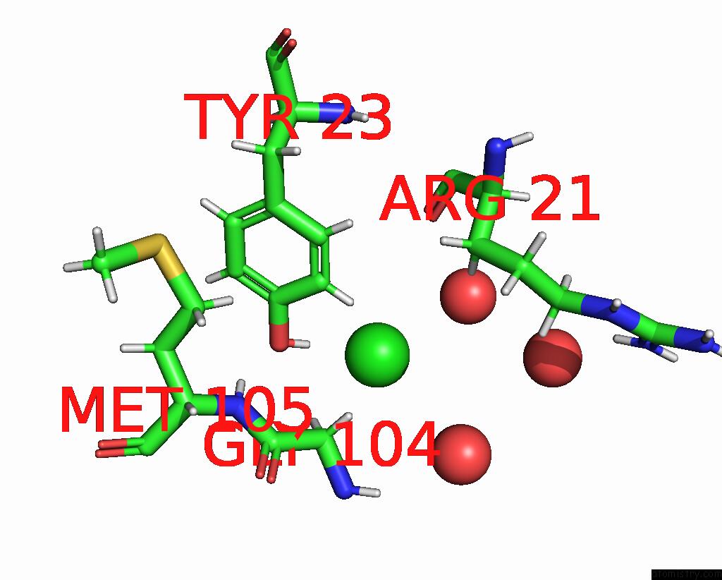
Chlorine binding site 3 out of 5 in 8cwd

Go back to Chlorine Binding Sites List in 8cwd
Chlorine binding site 3 out of 5 in the 20NS Temperature-Jump (DARK1) Xfel Structure of Lysozyme Bound to N, N'-Diacetylchitobiose


Mono view


Stereo pair view

A full contact list of Chlorine with other atoms in the Cl binding site number 3 of 20NS Temperature-Jump (DARK1) Xfel Structure of Lysozyme Bound to N, N'-Diacetylchitobiose within 5.0Å range:
probe atom residue distance (Å) B Occ
A:Cl206

b:20.5
occ:1.00
HH A:TYR23 2.1 16.6 1.0
OH A:TYR23 2.9 13.8 1.0
O A:HOH382 3.0 30.3 1.0
HE2 A:TYR23 3.0 15.2 1.0
HA3 A:GLY104 3.2 16.9 1.0
CE2 A:TYR23 3.5 12.7 1.0
CZ A:TYR23 3.6 12.7 1.0
HA2 A:GLY104 3.9 16.9 1.0
CA A:GLY104 4.0 14.1 1.0
O A:ARG21 4.4 21.2 1.0
O A:HOH305 4.4 28.3 1.0
N A:GLY104 4.5 15.9 1.0
H A:MET105 4.6 13.4 1.0
H A:GLY104 4.7 19.0 1.0
CD2 A:TYR23 4.7 11.7 1.0
CE1 A:TYR23 4.7 11.9 1.0
HB3 A:ARG21 4.8 26.0 1.0
O A:HOH358 4.9 29.1 1.0

Chlorine binding site 4 out of 5 in 8cwd

Go back to Chlorine Binding Sites List in 8cwd
Chlorine binding site 4 out of 5 in the 20NS Temperature-Jump (DARK1) Xfel Structure of Lysozyme Bound to N, N'-Diacetylchitobiose


Mono view


Stereo pair view

A full contact list of Chlorine with other atoms in the Cl binding site number 4 of 20NS Temperature-Jump (DARK1) Xfel Structure of Lysozyme Bound to N, N'-Diacetylchitobiose within 5.0Å range:
probe atom residue distance (Å) B Occ
A:Cl207

b:32.8
occ:1.00
H A:GLY26 2.2 14.6 1.0
HG3 A:GLN121 2.6 38.0 1.0
HA A:GLN121 2.7 22.8 1.0
HB2 A:SER24 2.8 21.2 1.0
HD13 A:ILE124 2.9 32.5 1.0
HE22 A:GLN121 2.9 41.4 1.0
HA3 A:GLY26 2.9 13.2 1.0
N A:GLY26 3.0 12.2 1.0
OG A:SER24 3.0 20.4 1.0
HG A:SER24 3.3 24.5 1.0
CB A:SER24 3.3 17.7 1.0
CG A:GLN121 3.4 31.7 1.0
HG23 A:VAL120 3.4 21.5 1.0
NE2 A:GLN121 3.4 34.6 1.0
CA A:GLY26 3.5 11.1 1.0
HG12 A:ILE124 3.5 27.5 1.0
CA A:GLN121 3.5 19.0 1.0
CD1 A:ILE124 3.7 27.1 1.0
CD A:GLN121 3.7 36.2 1.0
H A:LEU25 3.8 16.1 1.0
HD11 A:ILE124 3.8 32.5 1.0
N A:LEU25 3.9 13.4 1.0
N A:GLN121 3.9 17.5 1.0
CB A:GLN121 3.9 24.2 1.0
HE21 A:GLN121 4.0 41.4 1.0
HB3 A:SER24 4.0 21.2 1.0
C A:SER24 4.1 12.4 1.0
HA2 A:GLY26 4.1 13.2 1.0
HB3 A:LEU25 4.1 17.3 1.0
CG1 A:ILE124 4.1 22.9 1.0
H A:ASN27 4.1 12.7 1.0
HG2 A:GLN121 4.1 38.0 1.0
C A:LEU25 4.1 12.6 1.0
O A:VAL120 4.2 16.1 1.0
HB3 A:GLN121 4.2 29.1 1.0
H A:GLN121 4.3 21.0 1.0
C A:VAL120 4.3 15.6 1.0
CA A:SER24 4.3 14.9 1.0
CG2 A:VAL120 4.3 17.9 1.0
HD12 A:ILE124 4.5 32.5 1.0
HG23 A:ILE124 4.5 26.4 1.0
CA A:LEU25 4.5 13.7 1.0
C A:GLY26 4.5 11.2 1.0
HG21 A:VAL120 4.5 21.5 1.0
O A:SER24 4.6 13.2 1.0
N A:ASN27 4.6 10.6 1.0
HG13 A:ILE124 4.6 27.5 1.0
C A:GLN121 4.7 17.5 1.0
OE1 A:GLN121 4.8 37.6 1.0
HA A:SER24 4.8 17.9 1.0
HB2 A:GLN121 4.8 29.1 1.0
CB A:LEU25 4.8 14.4 1.0
HG22 A:VAL120 4.9 21.5 1.0
HB A:VAL120 4.9 19.7 1.0
O A:GLN121 4.9 20.1 1.0
HG21 A:ILE124 5.0 26.4 1.0

Chlorine binding site 5 out of 5 in 8cwd

Go back to Chlorine Binding Sites List in 8cwd
Chlorine binding site 5 out of 5 in the 20NS Temperature-Jump (DARK1) Xfel Structure of Lysozyme Bound to N, N'-Diacetylchitobiose


Mono view


Stereo pair view

A full contact list of Chlorine with other atoms in the Cl binding site number 5 of 20NS Temperature-Jump (DARK1) Xfel Structure of Lysozyme Bound to N, N'-Diacetylchitobiose within 5.0Å range:
probe atom residue distance (Å) B Occ
A:Cl208

b:113.7
occ:1.00
O A:HOH389 2.3 37.3 1.0
HZ1 A:LYS33 2.6 33.6 1.0
O A:HOH381 2.8 37.7 1.0
HZ2 A:LYS33 2.9 33.6 1.0
HE2 A:PHE38 3.1 18.6 1.0
NZ A:LYS33 3.2 28.0 1.0
HD2 A:PHE38 3.5 17.6 1.0
HZ3 A:LYS33 3.7 33.6 1.0
CE2 A:PHE38 3.8 15.5 1.0
O A:HOH316 3.8 25.2 1.0
HD3 A:LYS33 3.8 25.7 1.0
O A:HOH345 3.8 29.0 1.0
CD2 A:PHE38 4.0 14.7 1.0
CE A:LYS33 4.3 25.1 1.0
HB2 A:ARG5 4.3 21.4 1.0
CD A:LYS33 4.4 21.4 1.0
HE2 A:LYS33 4.5 30.1 1.0
HD2 A:LYS33 4.6 25.7 1.0
OD1 A:ASN37 4.8 24.0 0.9
HZ3 A:TRP123 4.9 26.9 1.0
H A:ARG5 4.9 20.1 1.0
CZ A:PHE38 4.9 14.5 1.0

Reference:

A.M.Wolff, E.Nango, I.D.Young, A.S.Brewster, M.Kubo, T.Nomura, M.Sugahara, S.Owada, B.A.Barad, K.Ito, A.Bhowmick, S.Carbajo, T.Hino, J.M.Holton, D.Im, L.J.O'riordan, T.Tanaka, R.Tanaka, R.G.Sierra, F.Yumoto, K.Tono, S.Iwata, N.K.Sauter, J.S.Fraser, M.C.Thompson. Mapping Protein Dynamics at High-Resolution with Temperature-Jump X-Ray Crystallography Biorxiv 2022.
DOI: 10.1101/2022.06.10.495662
Page generated: Sun Jul 13 10:25:07 2025

Last articles

Mg in 7KXG
Mg in 7KZ2
Mg in 7KZ4
Mg in 7KYB
Mg in 7KYC
Mg in 7KYA
Mg in 7KXH
Mg in 7KY9
Mg in 7KY8
Mg in 7KY7
© Copyright 2008-2020 by atomistry.com
Home   |    Site Map   |    Copyright   |    Contact us   |    Privacy