Chlorine »
PDB 8dcu-8dsi »
8dpv »
Chlorine in PDB 8dpv: The Crystal Structure of Interleukin-11, W147A Mutant
Protein crystallography data
The structure of The Crystal Structure of Interleukin-11, W147A Mutant, PDB code: 8dpv
was solved by
R.D.Metcalfe,
M.D.W.Griffin,
with X-Ray Crystallography technique. A brief refinement statistics is given in the table below:
| Resolution Low / High (Å) | 37.01 / 1.48 |
| Space group | P 21 21 2 |
| Cell size a, b, c (Å), α, β, γ (°) | 38.505, 133.996, 27.087, 90, 90, 90 |
| R / Rfree (%) | 18.3 / 21.6 |
Chlorine Binding Sites:
The binding sites of Chlorine atom in the The Crystal Structure of Interleukin-11, W147A Mutant
(pdb code 8dpv). This binding sites where shown within
5.0 Angstroms radius around Chlorine atom.
In total only one binding site of Chlorine was determined in the The Crystal Structure of Interleukin-11, W147A Mutant, PDB code: 8dpv:
In total only one binding site of Chlorine was determined in the The Crystal Structure of Interleukin-11, W147A Mutant, PDB code: 8dpv:
Chlorine binding site 1 out of 1 in 8dpv
Go back to
Chlorine binding site 1 out
of 1 in the The Crystal Structure of Interleukin-11, W147A Mutant
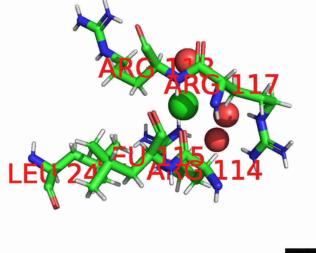
Mono view
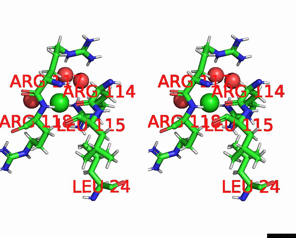
Stereo pair view
Mono view
Stereo pair view
A full contact list of Chlorine with other atoms in the Cl binding
site number 1 of The Crystal Structure of Interleukin-11, W147A Mutant within 5.0Å range:
|
Reference:
R.D.Metcalfe,
E.Hanssen,
K.Y.Fung,
K.Aizel,
C.C.Kosasih,
C.O.Zlatic,
L.Doughty,
C.J.Morton,
A.P.Leis,
M.W.Parker,
P.R.Gooley,
T.L.Putoczki,
M.D.W.Griffin.
Structures of the Interleukin 11 Signalling Complex Reveal GP130 Dynamics and the Inhibitory Mechanism of A Cytokine Variant Nat Commun V. 14 7543 2023.
ISSN: ESSN 2041-1723
DOI: 10.1038/S41467-023-42754-W
Page generated: Sun Jul 13 10:52:39 2025
ISSN: ESSN 2041-1723
DOI: 10.1038/S41467-023-42754-W
Last articles
Mn in 9LJUMn in 9LJW
Mn in 9LJS
Mn in 9LJR
Mn in 9LJT
Mn in 9LJV
Mg in 9UA2
Mg in 9R96
Mg in 9VM1
Mg in 9P01