Chlorine »
PDB 8ezt-8f55 »
8f10 »
Chlorine in PDB 8f10: Structure of the MDM2 P53 Binding Domain in Complex with H102, An All- D Helicon Polypeptide
Enzymatic activity of Structure of the MDM2 P53 Binding Domain in Complex with H102, An All- D Helicon Polypeptide
All present enzymatic activity of Structure of the MDM2 P53 Binding Domain in Complex with H102, An All- D Helicon Polypeptide:
2.3.2.27;
2.3.2.27;
Protein crystallography data
The structure of Structure of the MDM2 P53 Binding Domain in Complex with H102, An All- D Helicon Polypeptide, PDB code: 8f10
was solved by
K.Li,
A.J.Callahan,
T.L.Travaline,
O.S.Tokareva,
J.-M.Swiecicki,
G.L.Verdine,
B.L.Pentelute,
J.H.Mcgee,
with X-Ray Crystallography technique. A brief refinement statistics is given in the table below:
| Resolution Low / High (Å) | 28.02 / 1.28 |
| Space group | P 1 21 1 |
| Cell size a, b, c (Å), α, β, γ (°) | 28.15, 39.92, 42.36, 90, 95.44, 90 |
| R / Rfree (%) | 15.4 / 16.7 |
Chlorine Binding Sites:
The binding sites of Chlorine atom in the Structure of the MDM2 P53 Binding Domain in Complex with H102, An All- D Helicon Polypeptide
(pdb code 8f10). This binding sites where shown within
5.0 Angstroms radius around Chlorine atom.
In total only one binding site of Chlorine was determined in the Structure of the MDM2 P53 Binding Domain in Complex with H102, An All- D Helicon Polypeptide, PDB code: 8f10:
In total only one binding site of Chlorine was determined in the Structure of the MDM2 P53 Binding Domain in Complex with H102, An All- D Helicon Polypeptide, PDB code: 8f10:
Chlorine binding site 1 out of 1 in 8f10
Go back to
Chlorine binding site 1 out
of 1 in the Structure of the MDM2 P53 Binding Domain in Complex with H102, An All- D Helicon Polypeptide
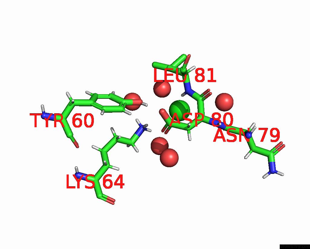
Mono view
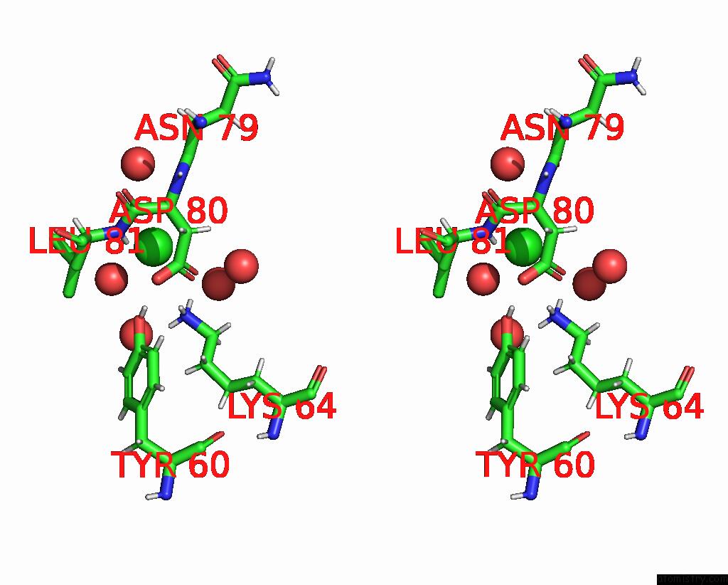
Stereo pair view
Mono view
Stereo pair view
A full contact list of Chlorine with other atoms in the Cl binding
site number 1 of Structure of the MDM2 P53 Binding Domain in Complex with H102, An All- D Helicon Polypeptide within 5.0Å range:
|
Reference:
A.J.Callahan,
S.Gandhesiri,
T.L.Travaline,
L.Lozano Salazar,
S.Hanna,
Y.-C.Lee,
K.Li,
O.S.Tokareva,
J.-M.Swiecicki,
A.Loas,
G.L.Verdine,
J.H.Mcgee,
B.L.Pentelute.
Single-Shot Flow Synthesis of D-Proteins For Mirror-Image Phage Display Chemrxiv 2023.
ISSN: ISSN 2573-2293
DOI: 10.26434/CHEMRXIV-2023-X86XP
Page generated: Sun Jul 13 11:10:01 2025
ISSN: ISSN 2573-2293
DOI: 10.26434/CHEMRXIV-2023-X86XP
Last articles
Fe in 9GYRFe in 9GYZ
Fe in 9GYU
Fe in 9GYY
Fe in 9GMC
Fe in 9GM3
Fe in 9GYD
Fe in 9G7I
Fe in 9GTJ
Fe in 9GS2