Chlorine »
PDB 8ezt-8f55 »
8f4u »
Chlorine in PDB 8f4u: Crystal Structure of Acetyltransferase Eis From M. Tuberculosis in Complex with Azelastine
Protein crystallography data
The structure of Crystal Structure of Acetyltransferase Eis From M. Tuberculosis in Complex with Azelastine, PDB code: 8f4u
was solved by
A.H.Pang,
A.Punetha,
S.Garneau-Tsodikova,
O.V.Tsodikov,
with X-Ray Crystallography technique. A brief refinement statistics is given in the table below:
| Resolution Low / High (Å) | 43.86 / 1.91 |
| Space group | H 3 2 |
| Cell size a, b, c (Å), α, β, γ (°) | 175.306, 175.306, 123.351, 90, 90, 120 |
| R / Rfree (%) | 16.7 / 19.5 |
Chlorine Binding Sites:
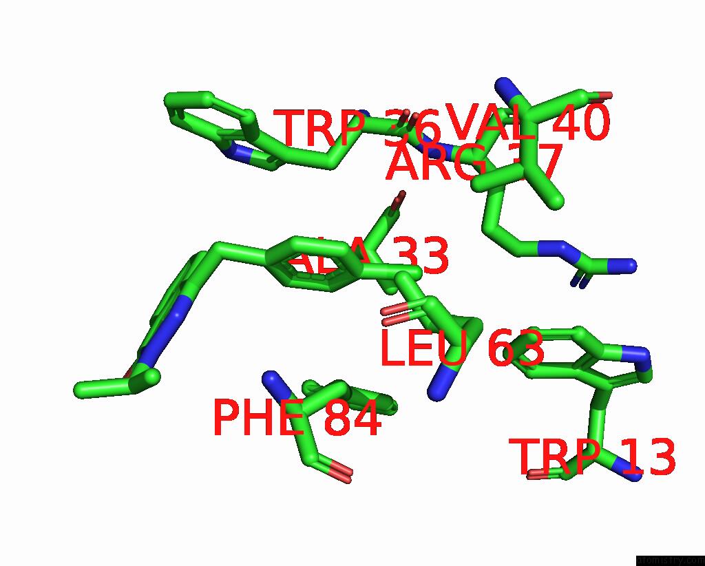
The binding sites of Chlorine atom in the Crystal Structure of Acetyltransferase Eis From M. Tuberculosis in Complex with Azelastine
(pdb code 8f4u). This binding sites where shown within
5.0 Angstroms radius around Chlorine atom.
In total only one binding site of Chlorine was determined in the Crystal Structure of Acetyltransferase Eis From M. Tuberculosis in Complex with Azelastine, PDB code: 8f4u:
In total only one binding site of Chlorine was determined in the Crystal Structure of Acetyltransferase Eis From M. Tuberculosis in Complex with Azelastine, PDB code: 8f4u:
Chlorine binding site 1 out of 1 in 8f4u
Go back to
Chlorine binding site 1 out
of 1 in the Crystal Structure of Acetyltransferase Eis From M. Tuberculosis in Complex with Azelastine
Mono view

Stereo pair view
Mono view
Stereo pair view
A full contact list of Chlorine with other atoms in the Cl binding
site number 1 of Crystal Structure of Acetyltransferase Eis From M. Tuberculosis in Complex with Azelastine within 5.0Å range:
|
Reference:
A.H.Pang,
K.D.Green,
A.Punetha,
N.Thamban Chandrika,
K.C.Howard,
S.Garneau-Tsodikova,
O.V.Tsodikov.
Discovery and Mechanistic Analysis of Structurally Diverse Inhibitors of Acetyltransferase Eis Among Fda-Approved Drugs. Biochemistry 2023.
ISSN: ISSN 0006-2960
PubMed: 36657084
DOI: 10.1021/ACS.BIOCHEM.2C00658
Page generated: Sun Jul 13 11:16:52 2025
ISSN: ISSN 0006-2960
PubMed: 36657084
DOI: 10.1021/ACS.BIOCHEM.2C00658
Last articles
Fe in 9GMCFe in 9GM3
Fe in 9GYD
Fe in 9G7I
Fe in 9GTJ
Fe in 9GS2
Fe in 9G9S
Fe in 9GK1
Fe in 9G03
Fe in 9G9R