Chlorine »
PDB 8gsg-8h8u »
8h7x »
Chlorine in PDB 8h7x: Crystal Structure of Egfr T790M/C797S Mutant in Complex with Brigatinib
Enzymatic activity of Crystal Structure of Egfr T790M/C797S Mutant in Complex with Brigatinib
All present enzymatic activity of Crystal Structure of Egfr T790M/C797S Mutant in Complex with Brigatinib:
2.7.10.1;
2.7.10.1;
Protein crystallography data
The structure of Crystal Structure of Egfr T790M/C797S Mutant in Complex with Brigatinib, PDB code: 8h7x
was solved by
M.Kukimoto-Niino,
M.Shirouzu,
with X-Ray Crystallography technique. A brief refinement statistics is given in the table below:
| Resolution Low / High (Å) | 47.95 / 3.40 |
| Space group | P 21 21 21 |
| Cell size a, b, c (Å), α, β, γ (°) | 50.1, 90.11, 165.31, 90, 90, 90 |
| R / Rfree (%) | 18.1 / 25.6 |
Chlorine Binding Sites:
The binding sites of Chlorine atom in the Crystal Structure of Egfr T790M/C797S Mutant in Complex with Brigatinib
(pdb code 8h7x). This binding sites where shown within
5.0 Angstroms radius around Chlorine atom.
In total only one binding site of Chlorine was determined in the Crystal Structure of Egfr T790M/C797S Mutant in Complex with Brigatinib, PDB code: 8h7x:
In total only one binding site of Chlorine was determined in the Crystal Structure of Egfr T790M/C797S Mutant in Complex with Brigatinib, PDB code: 8h7x:
Chlorine binding site 1 out of 1 in 8h7x
Go back to
Chlorine binding site 1 out
of 1 in the Crystal Structure of Egfr T790M/C797S Mutant in Complex with Brigatinib
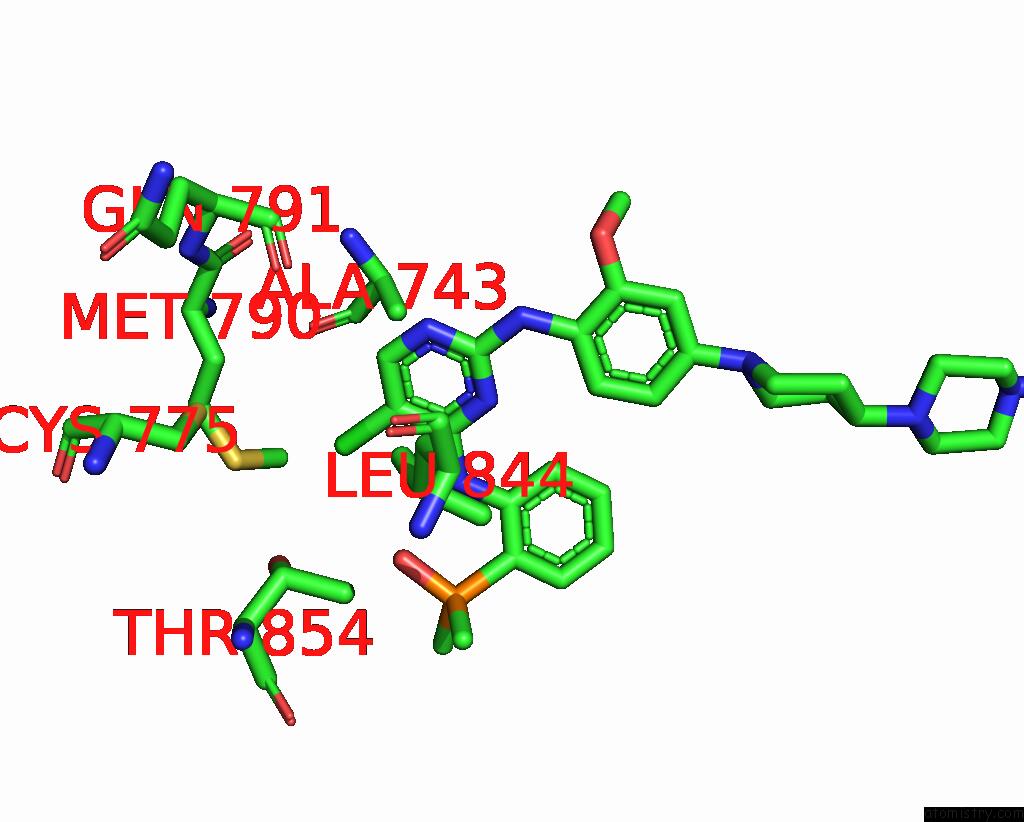
Mono view
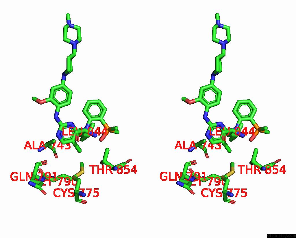
Stereo pair view
Mono view
Stereo pair view
A full contact list of Chlorine with other atoms in the Cl binding
site number 1 of Crystal Structure of Egfr T790M/C797S Mutant in Complex with Brigatinib within 5.0Å range:
|
Reference:
M.Suzuki,
K.Uchibori,
T.Oh-Hara,
Y.Nomura,
R.Suzuki,
M.Araki,
Y.Sagae,
M.Kukimoto-Niino,
S.Eguchi,
M.Shirouzu,
Y.Okuno,
N.Fujita,
R.Katayama.
Crystal Structure of Egfr T790M/C797S Mutant in Complex with Brigatinib To Be Published.
Page generated: Sun Jul 13 12:02:29 2025
Last articles
Mg in 7OUHMg in 7OUG
Mg in 7OUF
Mg in 7OU4
Mg in 7OU0
Mg in 7OTZ
Mg in 7OTP
Mg in 7OTX
Mg in 7OTO
Mg in 7OTJ