Chlorine »
PDB 8hwn-8ihz »
8i6b »
Chlorine in PDB 8i6b: Crystal Structure of Mycobacterium Tuberculosis Uracil-Dna Glycosylase in Complex with 5-Hydroxy-2,4(1H,3H)-Pyrimidinedione, Form I
Enzymatic activity of Crystal Structure of Mycobacterium Tuberculosis Uracil-Dna Glycosylase in Complex with 5-Hydroxy-2,4(1H,3H)-Pyrimidinedione, Form I
All present enzymatic activity of Crystal Structure of Mycobacterium Tuberculosis Uracil-Dna Glycosylase in Complex with 5-Hydroxy-2,4(1H,3H)-Pyrimidinedione, Form I:
3.2.2.27;
3.2.2.27;
Protein crystallography data
The structure of Crystal Structure of Mycobacterium Tuberculosis Uracil-Dna Glycosylase in Complex with 5-Hydroxy-2,4(1H,3H)-Pyrimidinedione, Form I, PDB code: 8i6b
was solved by
P.Raj,
A.Paul,
B.Gopal,
with X-Ray Crystallography technique. A brief refinement statistics is given in the table below:
| Resolution Low / High (Å) | 30.49 / 1.60 |
| Space group | P 1 21 1 |
| Cell size a, b, c (Å), α, β, γ (°) | 39.16, 63.68, 45.19, 90, 112.58, 90 |
| R / Rfree (%) | 14.9 / 18.2 |
Other elements in 8i6b:
The structure of Crystal Structure of Mycobacterium Tuberculosis Uracil-Dna Glycosylase in Complex with 5-Hydroxy-2,4(1H,3H)-Pyrimidinedione, Form I also contains other interesting chemical elements:
| Sodium | (Na) | 1 atom |
Chlorine Binding Sites:
The binding sites of Chlorine atom in the Crystal Structure of Mycobacterium Tuberculosis Uracil-Dna Glycosylase in Complex with 5-Hydroxy-2,4(1H,3H)-Pyrimidinedione, Form I
(pdb code 8i6b). This binding sites where shown within
5.0 Angstroms radius around Chlorine atom.
In total 3 binding sites of Chlorine where determined in the Crystal Structure of Mycobacterium Tuberculosis Uracil-Dna Glycosylase in Complex with 5-Hydroxy-2,4(1H,3H)-Pyrimidinedione, Form I, PDB code: 8i6b:
Jump to Chlorine binding site number: 1; 2; 3;
In total 3 binding sites of Chlorine where determined in the Crystal Structure of Mycobacterium Tuberculosis Uracil-Dna Glycosylase in Complex with 5-Hydroxy-2,4(1H,3H)-Pyrimidinedione, Form I, PDB code: 8i6b:
Jump to Chlorine binding site number: 1; 2; 3;
Chlorine binding site 1 out of 3 in 8i6b
Go back to
Chlorine binding site 1 out
of 3 in the Crystal Structure of Mycobacterium Tuberculosis Uracil-Dna Glycosylase in Complex with 5-Hydroxy-2,4(1H,3H)-Pyrimidinedione, Form I

Mono view
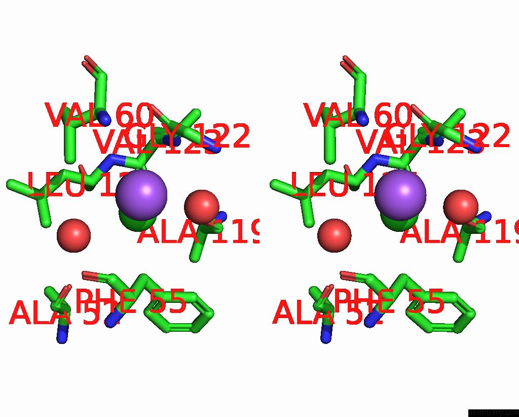
Stereo pair view
Mono view
Stereo pair view
A full contact list of Chlorine with other atoms in the Cl binding
site number 1 of Crystal Structure of Mycobacterium Tuberculosis Uracil-Dna Glycosylase in Complex with 5-Hydroxy-2,4(1H,3H)-Pyrimidinedione, Form I within 5.0Å range:
|
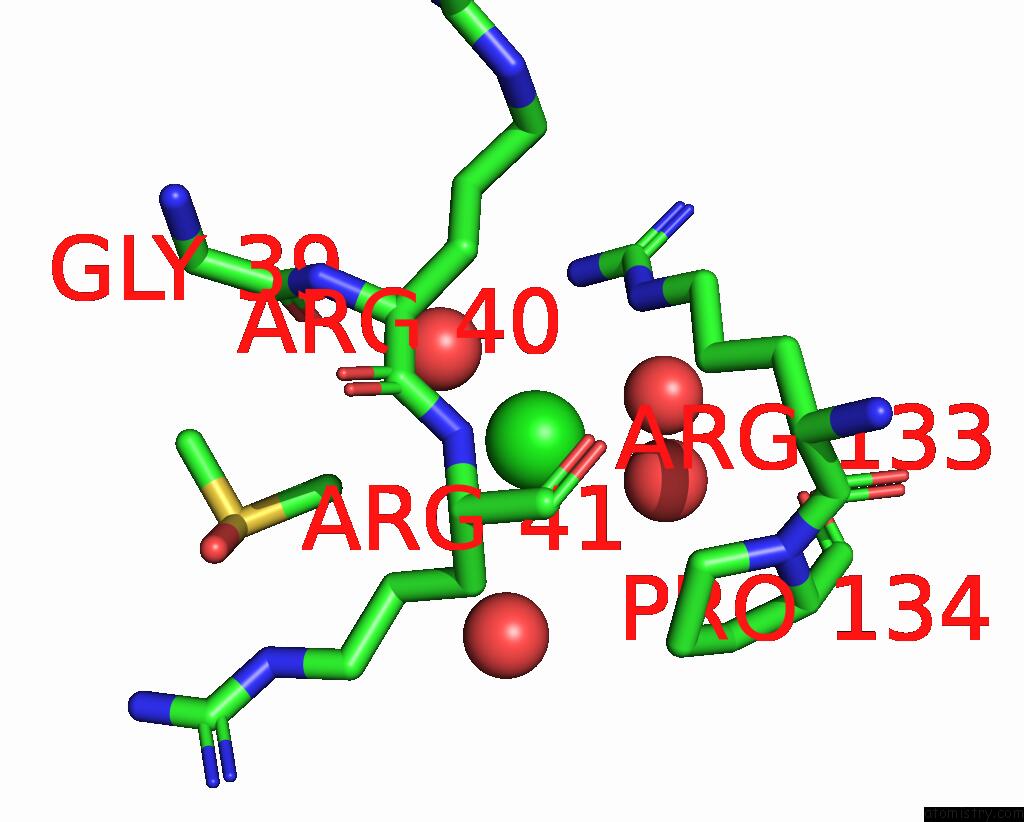
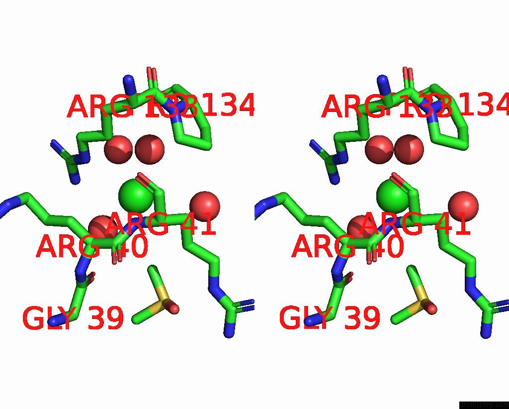
Chlorine binding site 2 out of 3 in 8i6b
Go back to
Chlorine binding site 2 out
of 3 in the Crystal Structure of Mycobacterium Tuberculosis Uracil-Dna Glycosylase in Complex with 5-Hydroxy-2,4(1H,3H)-Pyrimidinedione, Form I
Mono view
Stereo pair view
Mono view
Stereo pair view
A full contact list of Chlorine with other atoms in the Cl binding
site number 2 of Crystal Structure of Mycobacterium Tuberculosis Uracil-Dna Glycosylase in Complex with 5-Hydroxy-2,4(1H,3H)-Pyrimidinedione, Form I within 5.0Å range:
|
Chlorine binding site 3 out of 3 in 8i6b
Go back to
Chlorine binding site 3 out
of 3 in the Crystal Structure of Mycobacterium Tuberculosis Uracil-Dna Glycosylase in Complex with 5-Hydroxy-2,4(1H,3H)-Pyrimidinedione, Form I

Mono view
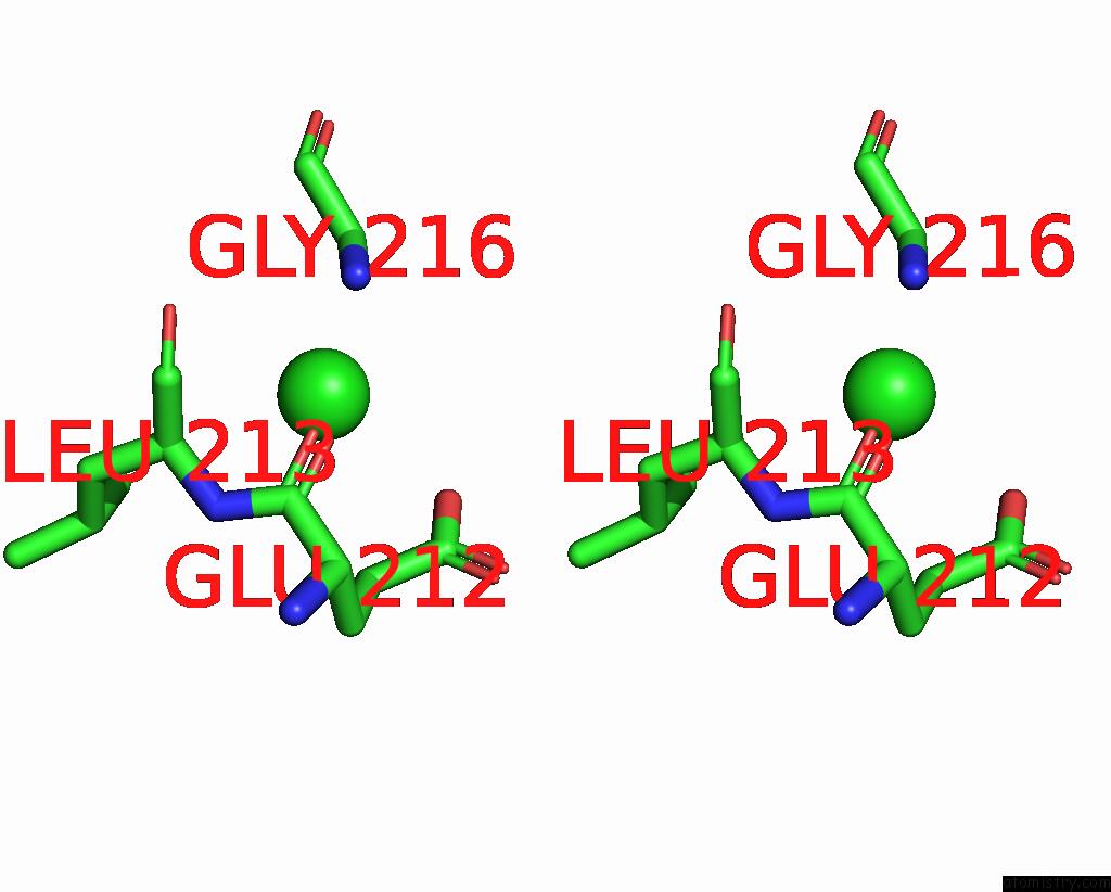
Stereo pair view
Mono view
Stereo pair view
A full contact list of Chlorine with other atoms in the Cl binding
site number 3 of Crystal Structure of Mycobacterium Tuberculosis Uracil-Dna Glycosylase in Complex with 5-Hydroxy-2,4(1H,3H)-Pyrimidinedione, Form I within 5.0Å range:
|
Reference:
S.Kesharwani,
P.Raj,
A.Paul,
K.Roy,
A.Bhanot,
A.Mehta,
A.Gopal,
U.Varshney,
B.Gopal,
S.Sundriyal.
Crystal Structures of Non-Uracil Ring Fragments in Complex with Mycobacterium Tuberculosis Uracil Dna Glycosylase (Mtung) As A Starting Point For Novel Inhibitor Design: A Case Study with the Barbituric Acid Fragment Eur.J.Med.Chem. V. 258 15604 2023.
ISSN: ISSN 0223-5234
DOI: 10.1016/J.EJMECH.2023.115604
Page generated: Sun Jul 13 12:11:14 2025
ISSN: ISSN 0223-5234
DOI: 10.1016/J.EJMECH.2023.115604
Last articles
Mg in 9EH5Mg in 9EI1
Mg in 9EHZ
Mg in 9EFU
Mg in 9EFG
Mg in 9ED3
Mg in 9ECO
Mg in 9E9Q
Mg in 9EB5
Mg in 9EAK