Chlorine »
PDB 8oez-8oqc »
8omw »
Chlorine in PDB 8omw: Crystal Structure of the Titin Domain FN3-20
Enzymatic activity of Crystal Structure of the Titin Domain FN3-20
All present enzymatic activity of Crystal Structure of the Titin Domain FN3-20:
2.7.11.1;
2.7.11.1;
Protein crystallography data
The structure of Crystal Structure of the Titin Domain FN3-20, PDB code: 8omw
was solved by
M.Rees,
M.Gautel,
with X-Ray Crystallography technique. A brief refinement statistics is given in the table below:
| Resolution Low / High (Å) | 31.09 / 1.05 |
| Space group | C 1 2 1 |
| Cell size a, b, c (Å), α, β, γ (°) | 49.034, 34.263, 63.081, 90, 99.68, 90 |
| R / Rfree (%) | 14.9 / 17.8 |
Chlorine Binding Sites:
The binding sites of Chlorine atom in the Crystal Structure of the Titin Domain FN3-20
(pdb code 8omw). This binding sites where shown within
5.0 Angstroms radius around Chlorine atom.
In total only one binding site of Chlorine was determined in the Crystal Structure of the Titin Domain FN3-20, PDB code: 8omw:
In total only one binding site of Chlorine was determined in the Crystal Structure of the Titin Domain FN3-20, PDB code: 8omw:
Chlorine binding site 1 out of 1 in 8omw
Go back to
Chlorine binding site 1 out
of 1 in the Crystal Structure of the Titin Domain FN3-20
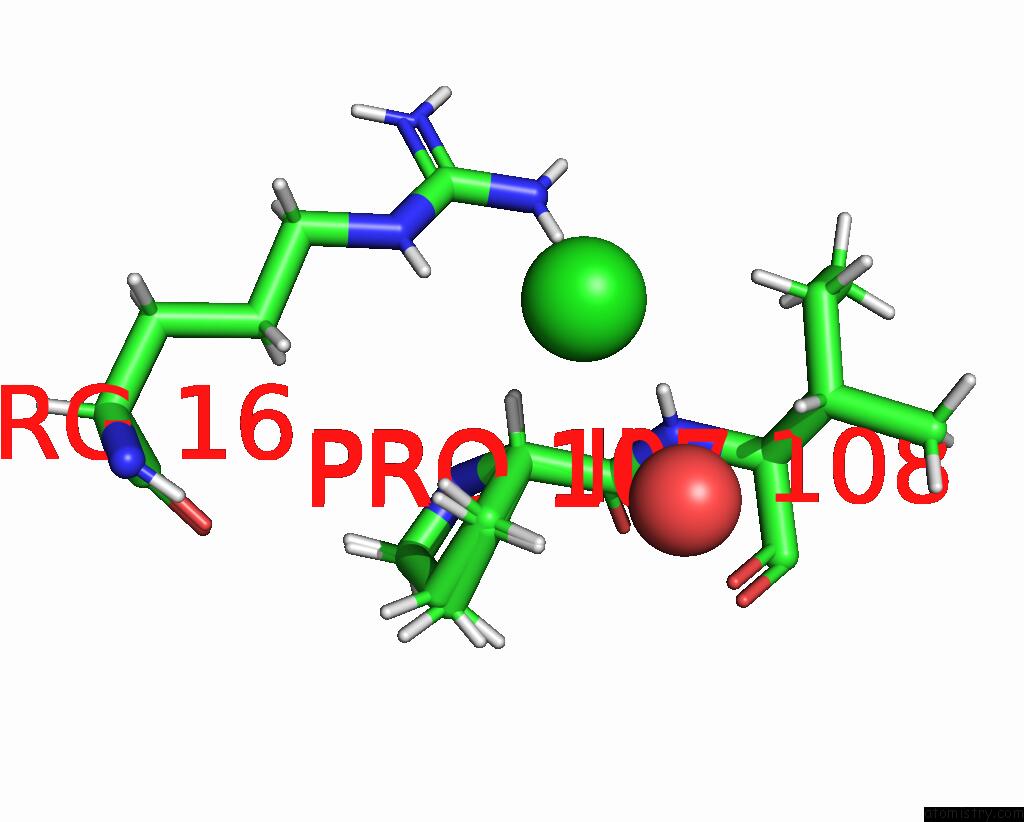
Mono view
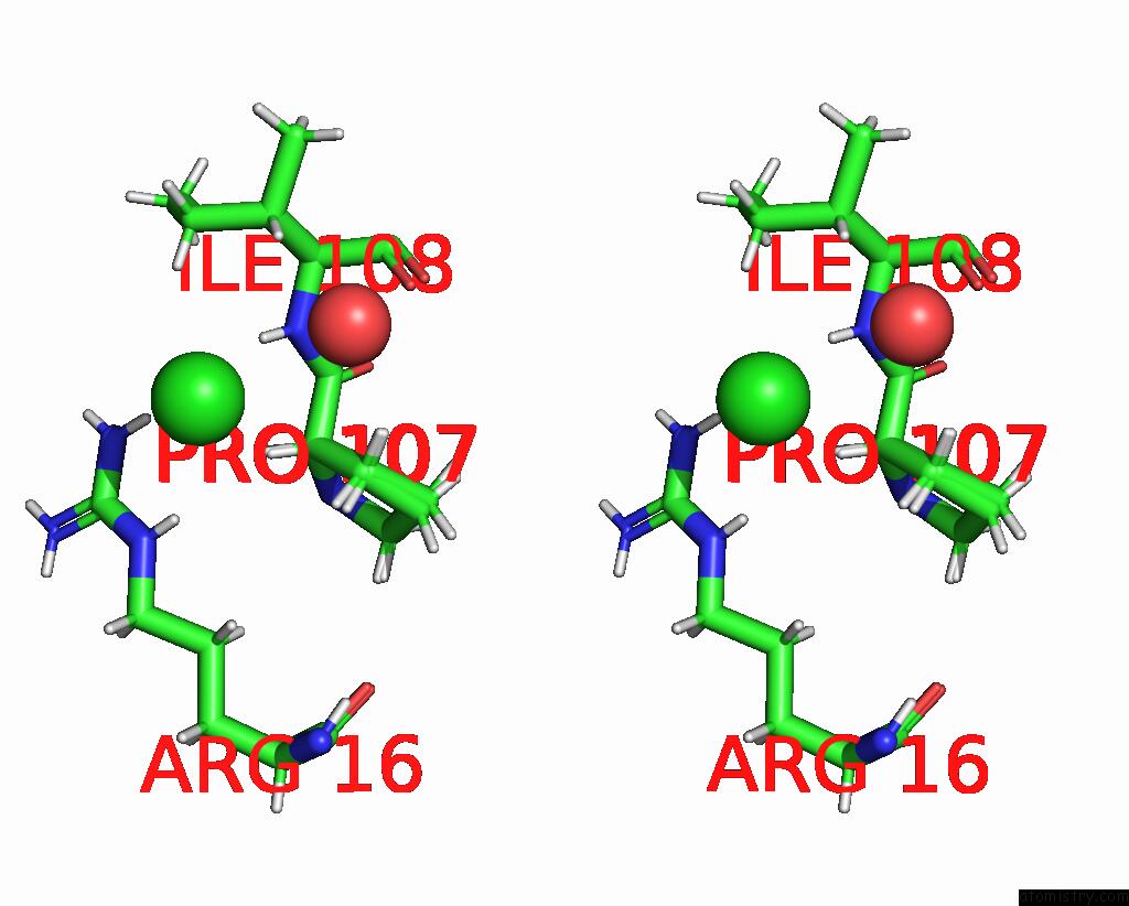
Stereo pair view
Mono view
Stereo pair view
A full contact list of Chlorine with other atoms in the Cl binding
site number 1 of Crystal Structure of the Titin Domain FN3-20 within 5.0Å range:
|
Reference:
M.Rees,
R.Nikoopour,
A.Alexandrovich,
M.Pfuhl,
L.R.Lopes,
M.M.Akhtar,
P.Syrris,
P.Elliott,
G.Carr-White,
M.Gautel.
Structure Determination and Analysis of Titin A-Band Fibronectin Type III Domains Provides Insights For Disease-Linked Variants and Protein Oligomerisation. J.Struct.Biol. 08009 2023.
ISSN: ESSN 1095-8657
PubMed: 37549721
DOI: 10.1016/J.JSB.2023.108009
Page generated: Sun Jul 13 12:51:24 2025
ISSN: ESSN 1095-8657
PubMed: 37549721
DOI: 10.1016/J.JSB.2023.108009
Last articles
Mg in 9FX7Mg in 9FX6
Mg in 9FWJ
Mg in 9FWA
Mg in 9FVJ
Mg in 9FVT
Mg in 9FTF
Mg in 9FU0
Mg in 9FSS
Mg in 9FS6