Chlorine »
PDB 8qlr-8qtf »
8qob »
Chlorine in PDB 8qob: Crystal Structure of Phosphoserine Phosphatase (Serb) From Brucella Melitensis in Complex with AP3 and Magnesium
Enzymatic activity of Crystal Structure of Phosphoserine Phosphatase (Serb) From Brucella Melitensis in Complex with AP3 and Magnesium
All present enzymatic activity of Crystal Structure of Phosphoserine Phosphatase (Serb) From Brucella Melitensis in Complex with AP3 and Magnesium:
3.1.3.3;
3.1.3.3;
Protein crystallography data
The structure of Crystal Structure of Phosphoserine Phosphatase (Serb) From Brucella Melitensis in Complex with AP3 and Magnesium, PDB code: 8qob
was solved by
T.Scaillet,
J.Wouters,
with X-Ray Crystallography technique. A brief refinement statistics is given in the table below:
| Resolution Low / High (Å) | 45.94 / 2.74 |
| Space group | I 21 3 |
| Cell size a, b, c (Å), α, β, γ (°) | 145.267, 145.267, 145.267, 90, 90, 90 |
| R / Rfree (%) | 18 / 24.3 |
Other elements in 8qob:
The structure of Crystal Structure of Phosphoserine Phosphatase (Serb) From Brucella Melitensis in Complex with AP3 and Magnesium also contains other interesting chemical elements:
| Magnesium | (Mg) | 1 atom |
Chlorine Binding Sites:
The binding sites of Chlorine atom in the Crystal Structure of Phosphoserine Phosphatase (Serb) From Brucella Melitensis in Complex with AP3 and Magnesium
(pdb code 8qob). This binding sites where shown within
5.0 Angstroms radius around Chlorine atom.
In total only one binding site of Chlorine was determined in the Crystal Structure of Phosphoserine Phosphatase (Serb) From Brucella Melitensis in Complex with AP3 and Magnesium, PDB code: 8qob:
In total only one binding site of Chlorine was determined in the Crystal Structure of Phosphoserine Phosphatase (Serb) From Brucella Melitensis in Complex with AP3 and Magnesium, PDB code: 8qob:
Chlorine binding site 1 out of 1 in 8qob
Go back to
Chlorine binding site 1 out
of 1 in the Crystal Structure of Phosphoserine Phosphatase (Serb) From Brucella Melitensis in Complex with AP3 and Magnesium
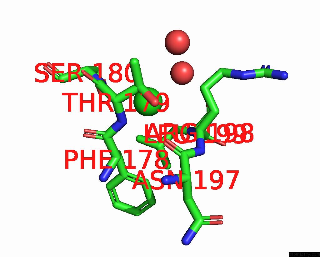
Mono view
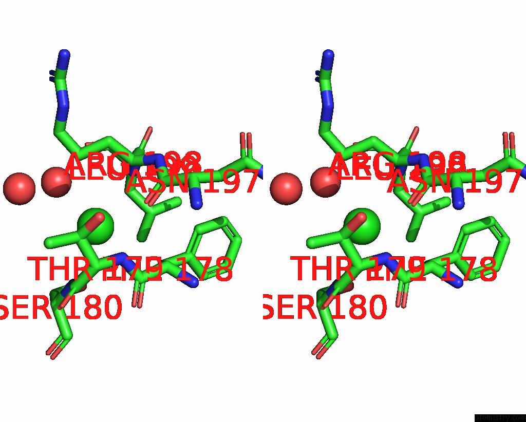
Stereo pair view
Mono view
Stereo pair view
A full contact list of Chlorine with other atoms in the Cl binding
site number 1 of Crystal Structure of Phosphoserine Phosphatase (Serb) From Brucella Melitensis in Complex with AP3 and Magnesium within 5.0Å range:
|
Reference:
T.Scaillet,
J.Wouters.
Crystal Structure of Phosphoserine Phosphatase (Serb) From Brucella Melitensis in Complex with AP3 and Magnesium To Be Published.
Page generated: Sun Jul 13 13:37:13 2025
Last articles
Mg in 8WTOMg in 8WT9
Mg in 8WTL
Mg in 8WTK
Mg in 8WT7
Mg in 8WT8
Mg in 8WT6
Mg in 8WR4
Mg in 8WT0
Mg in 8WSM