Chlorine »
PDB 8qjo-8qsc »
8qq5 »
Chlorine in PDB 8qq5: Structure of Wt Spnox Dh Domain: A Bacterial Nadph Oxidase.
Protein crystallography data
The structure of Structure of Wt Spnox Dh Domain: A Bacterial Nadph Oxidase., PDB code: 8qq5
was solved by
M.Thepaut,
I.Petit-Hartlein,
A.Vermot,
A.S.Humm,
F.Dupeux,
J.A.Marquez,
S.Smith,
F.Fieschi,
with X-Ray Crystallography technique. A brief refinement statistics is given in the table below:
| Resolution Low / High (Å) | 46.90 / 2.50 |
| Space group | P 41 21 2 |
| Cell size a, b, c (Å), α, β, γ (°) | 104.88, 104.88, 139.29, 90, 90, 90 |
| R / Rfree (%) | 19.7 / 29 |
Chlorine Binding Sites:
The binding sites of Chlorine atom in the Structure of Wt Spnox Dh Domain: A Bacterial Nadph Oxidase.
(pdb code 8qq5). This binding sites where shown within
5.0 Angstroms radius around Chlorine atom.
In total 2 binding sites of Chlorine where determined in the Structure of Wt Spnox Dh Domain: A Bacterial Nadph Oxidase., PDB code: 8qq5:
Jump to Chlorine binding site number: 1; 2;
In total 2 binding sites of Chlorine where determined in the Structure of Wt Spnox Dh Domain: A Bacterial Nadph Oxidase., PDB code: 8qq5:
Jump to Chlorine binding site number: 1; 2;
Chlorine binding site 1 out of 2 in 8qq5
Go back to
Chlorine binding site 1 out
of 2 in the Structure of Wt Spnox Dh Domain: A Bacterial Nadph Oxidase.
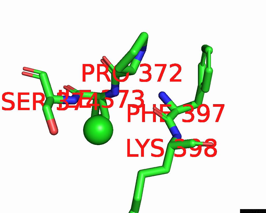
Mono view
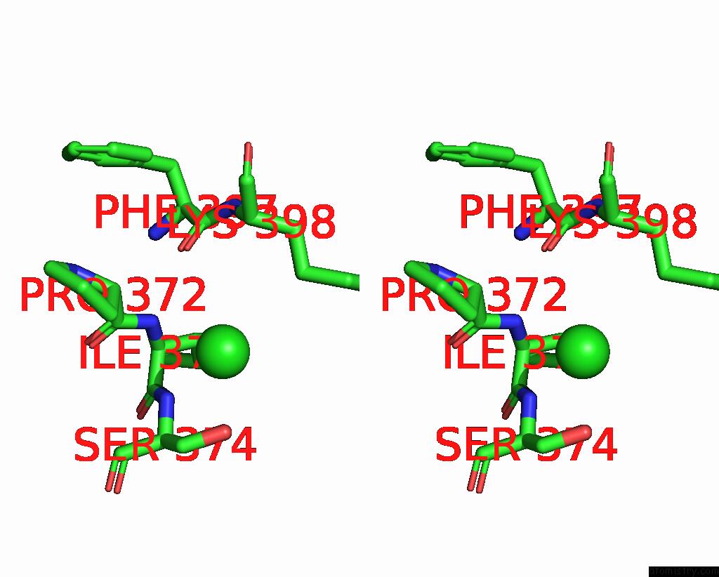
Stereo pair view
Mono view
Stereo pair view
A full contact list of Chlorine with other atoms in the Cl binding
site number 1 of Structure of Wt Spnox Dh Domain: A Bacterial Nadph Oxidase. within 5.0Å range:
|
Chlorine binding site 2 out of 2 in 8qq5
Go back to
Chlorine binding site 2 out
of 2 in the Structure of Wt Spnox Dh Domain: A Bacterial Nadph Oxidase.
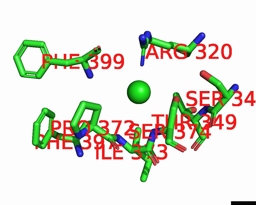
Mono view
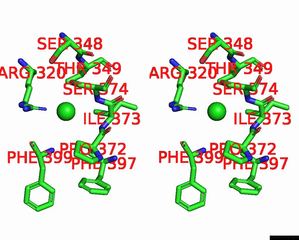
Stereo pair view
Mono view
Stereo pair view
A full contact list of Chlorine with other atoms in the Cl binding
site number 2 of Structure of Wt Spnox Dh Domain: A Bacterial Nadph Oxidase. within 5.0Å range:
|
Reference:
I.Petit-Hartlein,
A.Vermot,
M.Thepaut,
A.S.Humm,
F.Dupeux,
J.Dupuy,
V.Chaptal,
J.A.Marquez,
S.M.E.Smith,
F.Fieschi.
X-Ray Structure and Enzymatic Study of A Bacterial Nadph Oxidase Highlight the Activation Mechanism of Eukaryotic Nox. Elife V. 13 2024.
ISSN: ESSN 2050-084X
PubMed: 38640072
DOI: 10.7554/ELIFE.93759
Page generated: Sun Jul 13 13:37:57 2025
ISSN: ESSN 2050-084X
PubMed: 38640072
DOI: 10.7554/ELIFE.93759
Last articles
Mn in 9LJUMn in 9LJW
Mn in 9LJS
Mn in 9LJR
Mn in 9LJT
Mn in 9LJV
Mg in 9UA2
Mg in 9R96
Mg in 9VM1
Mg in 9P01