Chlorine »
PDB 8r67-8riu »
8rb1 »
Chlorine in PDB 8rb1: The Crystal Structure of Dna-Bound Human Mutsbeta (MSH2_K675R/MSH3_K902R) in the Canonical Mismatch Bound Conformation with Adp Bound in MSH2 and MSH3
Protein crystallography data
The structure of The Crystal Structure of Dna-Bound Human Mutsbeta (MSH2_K675R/MSH3_K902R) in the Canonical Mismatch Bound Conformation with Adp Bound in MSH2 and MSH3, PDB code: 8rb1
was solved by
M.Thomsen,
E.Costanzi,
with X-Ray Crystallography technique. A brief refinement statistics is given in the table below:
| Resolution Low / High (Å) | 86.66 / 2.09 |
| Space group | P 1 |
| Cell size a, b, c (Å), α, β, γ (°) | 69.705, 91.681, 94.024, 67.34, 87.16, 75.86 |
| R / Rfree (%) | 19.8 / 23.5 |
Other elements in 8rb1:
The structure of The Crystal Structure of Dna-Bound Human Mutsbeta (MSH2_K675R/MSH3_K902R) in the Canonical Mismatch Bound Conformation with Adp Bound in MSH2 and MSH3 also contains other interesting chemical elements:
| Magnesium | (Mg) | 2 atoms |
Chlorine Binding Sites:
The binding sites of Chlorine atom in the The Crystal Structure of Dna-Bound Human Mutsbeta (MSH2_K675R/MSH3_K902R) in the Canonical Mismatch Bound Conformation with Adp Bound in MSH2 and MSH3
(pdb code 8rb1). This binding sites where shown within
5.0 Angstroms radius around Chlorine atom.
In total only one binding site of Chlorine was determined in the The Crystal Structure of Dna-Bound Human Mutsbeta (MSH2_K675R/MSH3_K902R) in the Canonical Mismatch Bound Conformation with Adp Bound in MSH2 and MSH3, PDB code: 8rb1:
In total only one binding site of Chlorine was determined in the The Crystal Structure of Dna-Bound Human Mutsbeta (MSH2_K675R/MSH3_K902R) in the Canonical Mismatch Bound Conformation with Adp Bound in MSH2 and MSH3, PDB code: 8rb1:
Chlorine binding site 1 out of 1 in 8rb1
Go back to
Chlorine binding site 1 out
of 1 in the The Crystal Structure of Dna-Bound Human Mutsbeta (MSH2_K675R/MSH3_K902R) in the Canonical Mismatch Bound Conformation with Adp Bound in MSH2 and MSH3
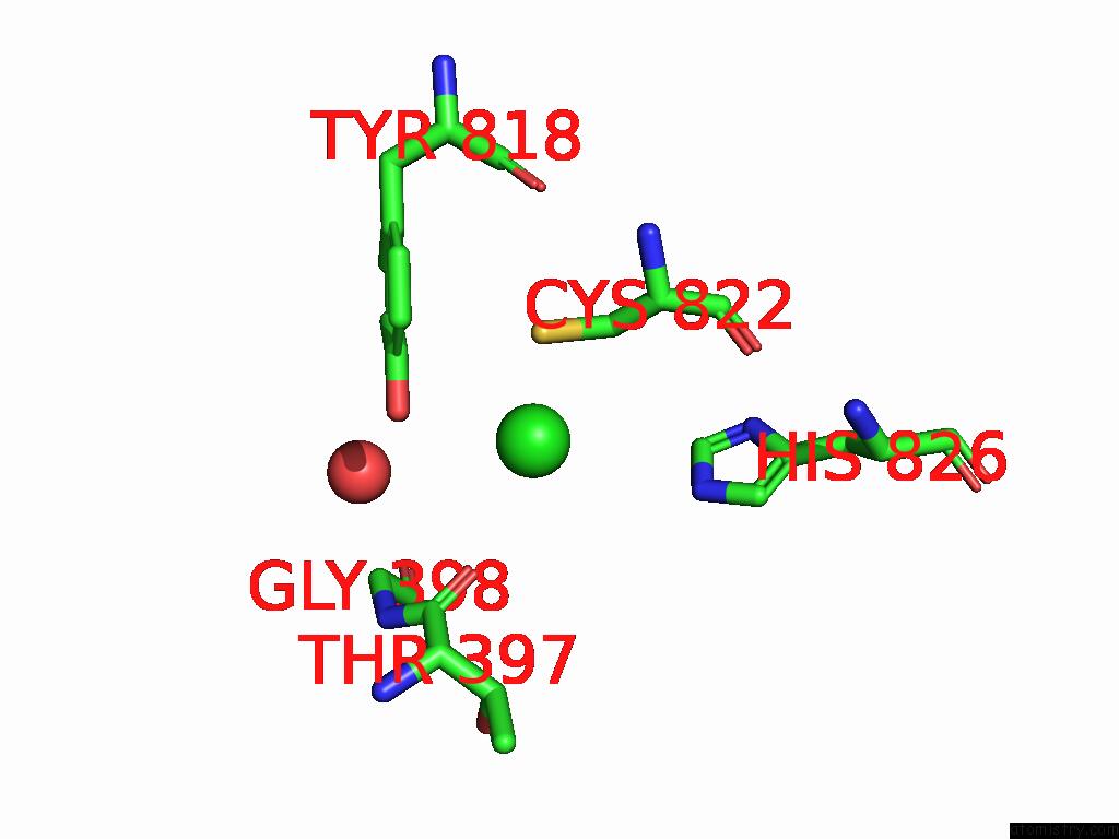
Mono view
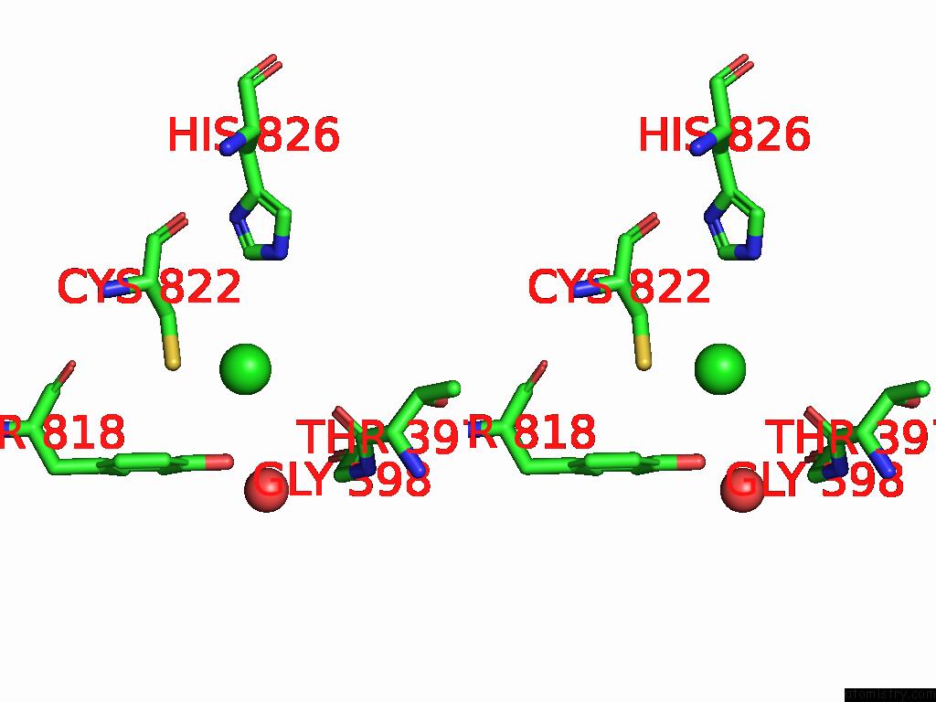
Stereo pair view
Mono view
Stereo pair view
A full contact list of Chlorine with other atoms in the Cl binding
site number 1 of The Crystal Structure of Dna-Bound Human Mutsbeta (MSH2_K675R/MSH3_K902R) in the Canonical Mismatch Bound Conformation with Adp Bound in MSH2 and MSH3 within 5.0Å range:
|
Reference:
M.Thomsen,
T.Neudegger,
G.Thieulin-Pardo,
M.Blaesse,
E.Costanzi,
S.Steinbacher,
N.V.Plotnikov,
C.Dominguez,
R.R.Iyer,
H.A.Wilkinson,
E.Monteagudo,
T.S.Haque,
B.C.Prasad,
M.Finley,
J.Boudet,
T.F.Vogt,
D.P.Felsenfeld.
The Crystal Structure of Dna-Bound Human Mutsbeta (MSH2/MSH3) in the Canonical Mismatch Bound Conformation with Adp Bound in MSH2 and MSH3 To Be Published.
Page generated: Sun Jul 13 13:52:11 2025
Last articles
Mg in 8OSFMg in 8OUK
Mg in 8OU0
Mg in 8OSG
Mg in 8OTV
Mg in 8OSO
Mg in 8OSN
Mg in 8OSM
Mg in 8OOI
Mg in 8ORJ