Chlorine »
PDB 8rx0-8sd4 »
8s5a »
Chlorine in PDB 8s5a: The Crystal Structure of FAN1 Nuclease Bound to 5' Phosphorylated P(Dg)/3'(Dt-Dt-Dt-Dt) Double Flap Dna
Enzymatic activity of The Crystal Structure of FAN1 Nuclease Bound to 5' Phosphorylated P(Dg)/3'(Dt-Dt-Dt-Dt) Double Flap Dna
All present enzymatic activity of The Crystal Structure of FAN1 Nuclease Bound to 5' Phosphorylated P(Dg)/3'(Dt-Dt-Dt-Dt) Double Flap Dna:
3.1.4.1;
3.1.4.1;
Protein crystallography data
The structure of The Crystal Structure of FAN1 Nuclease Bound to 5' Phosphorylated P(Dg)/3'(Dt-Dt-Dt-Dt) Double Flap Dna, PDB code: 8s5a
was solved by
E.Costanzi,
M.Thomsen,
with X-Ray Crystallography technique. A brief refinement statistics is given in the table below:
| Resolution Low / High (Å) | 43.40 / 2.65 |
| Space group | P 21 21 21 |
| Cell size a, b, c (Å), α, β, γ (°) | 91.626, 100.547, 112.879, 90, 90, 90 |
| R / Rfree (%) | 23.4 / 27.3 |
Other elements in 8s5a:
The structure of The Crystal Structure of FAN1 Nuclease Bound to 5' Phosphorylated P(Dg)/3'(Dt-Dt-Dt-Dt) Double Flap Dna also contains other interesting chemical elements:
| Calcium | (Ca) | 2 atoms |
Chlorine Binding Sites:
The binding sites of Chlorine atom in the The Crystal Structure of FAN1 Nuclease Bound to 5' Phosphorylated P(Dg)/3'(Dt-Dt-Dt-Dt) Double Flap Dna
(pdb code 8s5a). This binding sites where shown within
5.0 Angstroms radius around Chlorine atom.
In total only one binding site of Chlorine was determined in the The Crystal Structure of FAN1 Nuclease Bound to 5' Phosphorylated P(Dg)/3'(Dt-Dt-Dt-Dt) Double Flap Dna, PDB code: 8s5a:
In total only one binding site of Chlorine was determined in the The Crystal Structure of FAN1 Nuclease Bound to 5' Phosphorylated P(Dg)/3'(Dt-Dt-Dt-Dt) Double Flap Dna, PDB code: 8s5a:
Chlorine binding site 1 out of 1 in 8s5a
Go back to
Chlorine binding site 1 out
of 1 in the The Crystal Structure of FAN1 Nuclease Bound to 5' Phosphorylated P(Dg)/3'(Dt-Dt-Dt-Dt) Double Flap Dna
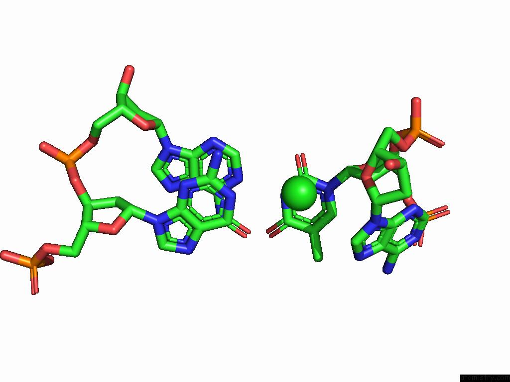
Mono view
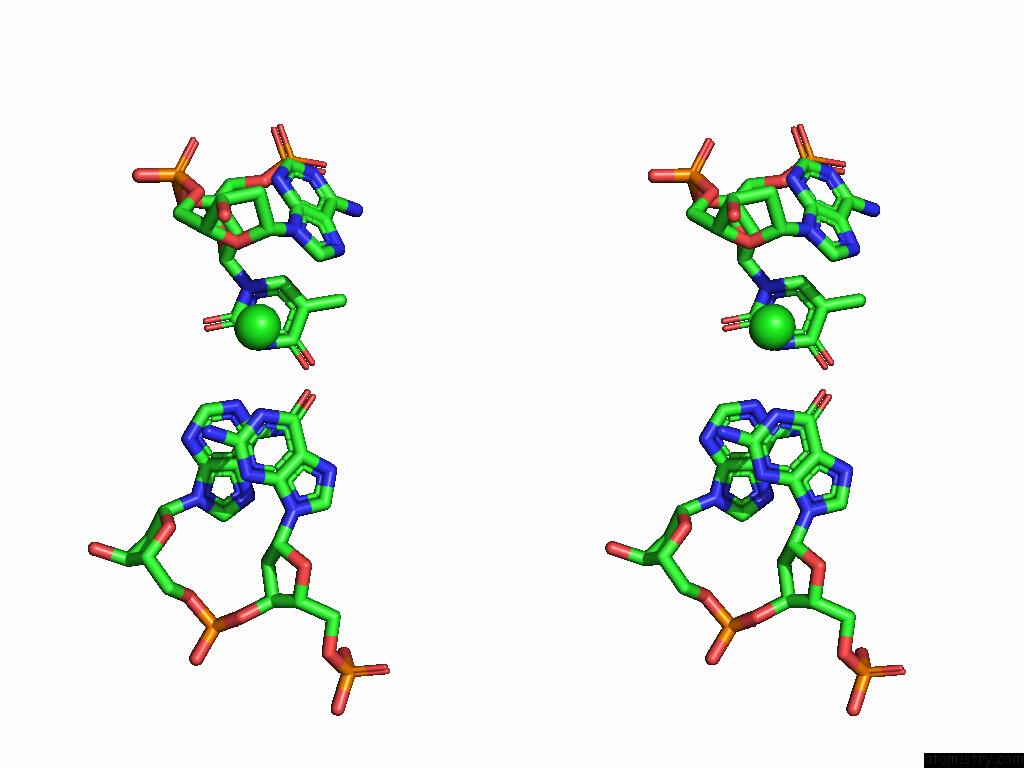
Stereo pair view
Mono view
Stereo pair view
A full contact list of Chlorine with other atoms in the Cl binding
site number 1 of The Crystal Structure of FAN1 Nuclease Bound to 5' Phosphorylated P(Dg)/3'(Dt-Dt-Dt-Dt) Double Flap Dna within 5.0Å range:
|
Reference:
E.Costanzi,
M.Thomsen.
The Crystal Structure of FAN1 Nuclease Bound to 5' Phosphorylated P(Dg)/3'(Dt-Dt-Dt-Dt) Double Flap Dna To Be Published.
Page generated: Sun Jul 13 14:03:24 2025
Last articles
Mg in 4IR7Mg in 4IQY
Mg in 4IPE
Mg in 4IP2
Mg in 4IP5
Mg in 4IP4
Mg in 4IOA
Mg in 4IOK
Mg in 4IOC
Mg in 4IO9