Chlorine »
PDB 8tgx-8tv1 »
8tms »
Chlorine in PDB 8tms: Crystal Structure of Bacterial Pectin Methylesterase PMEC2 From Rumen Butyrivibrio
Protein crystallography data
The structure of Crystal Structure of Bacterial Pectin Methylesterase PMEC2 From Rumen Butyrivibrio, PDB code: 8tms
was solved by
V.Carbone,
K.Reilly,
C.Sang,
L.Schofield,
R.Ronimus,
W.J.Kelly,
G.T.Attwood,
N.Palevich,
with X-Ray Crystallography technique. A brief refinement statistics is given in the table below:
| Resolution Low / High (Å) | 47.17 / 2.30 |
| Space group | P 1 |
| Cell size a, b, c (Å), α, β, γ (°) | 48.638, 76.282, 96.779, 98.36, 104.16, 90.05 |
| R / Rfree (%) | 20.4 / 24 |
Chlorine Binding Sites:
The binding sites of Chlorine atom in the Crystal Structure of Bacterial Pectin Methylesterase PMEC2 From Rumen Butyrivibrio
(pdb code 8tms). This binding sites where shown within
5.0 Angstroms radius around Chlorine atom.
In total 2 binding sites of Chlorine where determined in the Crystal Structure of Bacterial Pectin Methylesterase PMEC2 From Rumen Butyrivibrio, PDB code: 8tms:
Jump to Chlorine binding site number: 1; 2;
In total 2 binding sites of Chlorine where determined in the Crystal Structure of Bacterial Pectin Methylesterase PMEC2 From Rumen Butyrivibrio, PDB code: 8tms:
Jump to Chlorine binding site number: 1; 2;
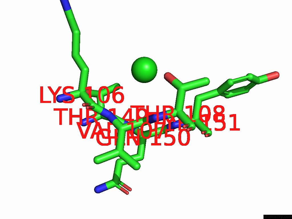
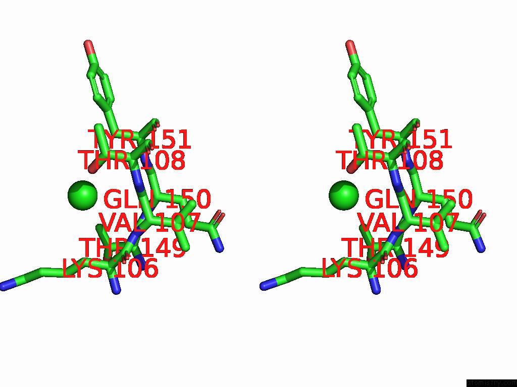
Chlorine binding site 1 out of 2 in 8tms
Go back to
Chlorine binding site 1 out
of 2 in the Crystal Structure of Bacterial Pectin Methylesterase PMEC2 From Rumen Butyrivibrio

Mono view

Stereo pair view
Mono view
Stereo pair view
A full contact list of Chlorine with other atoms in the Cl binding
site number 1 of Crystal Structure of Bacterial Pectin Methylesterase PMEC2 From Rumen Butyrivibrio within 5.0Å range:
|
Chlorine binding site 2 out of 2 in 8tms
Go back to
Chlorine binding site 2 out
of 2 in the Crystal Structure of Bacterial Pectin Methylesterase PMEC2 From Rumen Butyrivibrio
Mono view
Stereo pair view
Mono view
Stereo pair view
A full contact list of Chlorine with other atoms in the Cl binding
site number 2 of Crystal Structure of Bacterial Pectin Methylesterase PMEC2 From Rumen Butyrivibrio within 5.0Å range:
|
Reference:
V.Carbone,
K.Reilly,
C.Sang,
L.Schofield,
R.Ronimus,
W.J.Kelly,
G.T.Attwood,
N.Palevich.
Crystal Structures of Bacterial Pectin Methylesterases PME8A and PMEC2 From Rumen Butyrivibrio To Be Published.
Page generated: Sun Jul 13 14:29:38 2025
Last articles
K in 2QBMK in 2QBL
K in 2Q8I
K in 2Q8H
K in 2Q01
K in 2Q8G
K in 2Q8F
K in 2Q3V
K in 2PGA
K in 2PMU