Chlorine »
PDB 8uou-8uz7 »
8uuc »
Chlorine in PDB 8uuc: Crystal Structure of A Bacterial Clusterless Mutyx Bound to An Abasic Site Analog (Thf) Opposite D(8-Oxo-G)
Protein crystallography data
The structure of Crystal Structure of A Bacterial Clusterless Mutyx Bound to An Abasic Site Analog (Thf) Opposite D(8-Oxo-G), PDB code: 8uuc
was solved by
C.H.Trasvina-Arenas,
S.S.David,
A.J.Fisher,
with X-Ray Crystallography technique. A brief refinement statistics is given in the table below:
| Resolution Low / High (Å) | 37.62 / 1.55 |
| Space group | C 2 2 21 |
| Cell size a, b, c (Å), α, β, γ (°) | 38.832, 84.137, 225.742, 90, 90, 90 |
| R / Rfree (%) | 17.6 / 19.7 |
Other elements in 8uuc:
The structure of Crystal Structure of A Bacterial Clusterless Mutyx Bound to An Abasic Site Analog (Thf) Opposite D(8-Oxo-G) also contains other interesting chemical elements:
| Magnesium | (Mg) | 2 atoms |
| Sodium | (Na) | 2 atoms |
Chlorine Binding Sites:
The binding sites of Chlorine atom in the Crystal Structure of A Bacterial Clusterless Mutyx Bound to An Abasic Site Analog (Thf) Opposite D(8-Oxo-G)
(pdb code 8uuc). This binding sites where shown within
5.0 Angstroms radius around Chlorine atom.
In total only one binding site of Chlorine was determined in the Crystal Structure of A Bacterial Clusterless Mutyx Bound to An Abasic Site Analog (Thf) Opposite D(8-Oxo-G), PDB code: 8uuc:
In total only one binding site of Chlorine was determined in the Crystal Structure of A Bacterial Clusterless Mutyx Bound to An Abasic Site Analog (Thf) Opposite D(8-Oxo-G), PDB code: 8uuc:
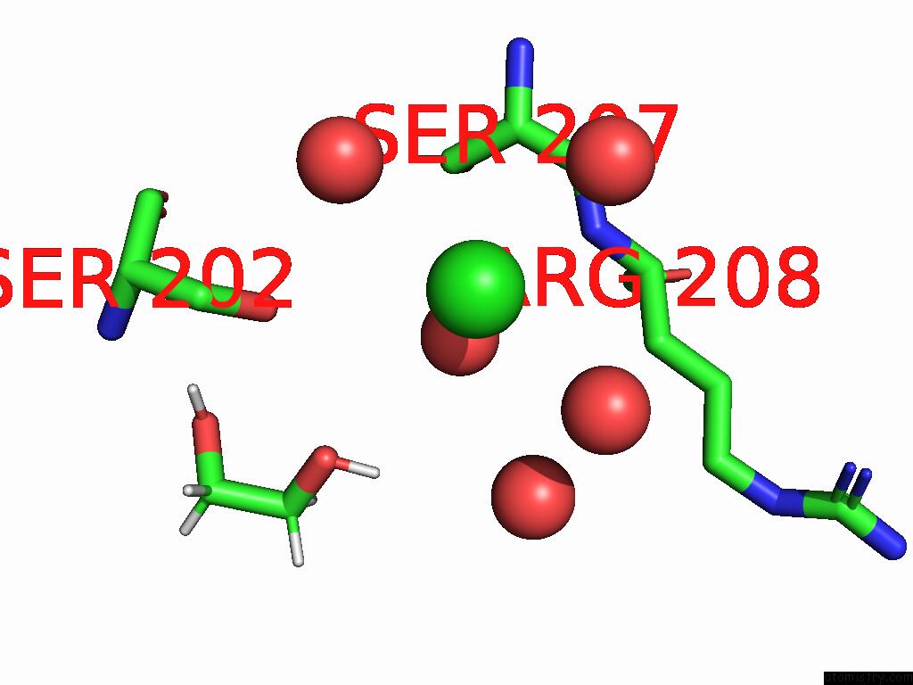
Chlorine binding site 1 out of 1 in 8uuc
Go back to
Chlorine binding site 1 out
of 1 in the Crystal Structure of A Bacterial Clusterless Mutyx Bound to An Abasic Site Analog (Thf) Opposite D(8-Oxo-G)
Mono view

Stereo pair view
Mono view
Stereo pair view
A full contact list of Chlorine with other atoms in the Cl binding
site number 1 of Crystal Structure of A Bacterial Clusterless Mutyx Bound to An Abasic Site Analog (Thf) Opposite D(8-Oxo-G) within 5.0Å range:
|
Reference:
C.H.Trasvina-Arenas,
M.Hashemian,
M.Malek,
S.Merrill,
A.J.Fisher,
S.S.David.
Crystal Structure of Mutyx: A Novel Clusterless Adenine Dna Glycosylase with A Distinct C-Terminal Domain and 8-Oxoguanine Recognition Sphere. Biorxiv 2025.
ISSN: ISSN 2692-8205
PubMed: 39803464
DOI: 10.1101/2025.01.03.631205
Page generated: Sun Jul 13 15:06:19 2025
ISSN: ISSN 2692-8205
PubMed: 39803464
DOI: 10.1101/2025.01.03.631205
Last articles
Mg in 4WF7Mg in 4WEC
Mg in 4WEO
Mg in 4WCW
Mg in 4WE1
Mg in 4WCU
Mg in 4WDQ
Mg in 4WB6
Mg in 4WB8
Mg in 4WC0