Chlorine »
PDB 8xet-8yq4 »
8xfi »
Chlorine in PDB 8xfi: High-Resolution Structure of the Siderophore Periplasmic Binding Protein Ftsb From Streptococcus Pyogenes with Ferrioxamine E Bound (Crystal Form 2)
Protein crystallography data
The structure of High-Resolution Structure of the Siderophore Periplasmic Binding Protein Ftsb From Streptococcus Pyogenes with Ferrioxamine E Bound (Crystal Form 2), PDB code: 8xfi
was solved by
J.M.M.Caaveiro,
J.Fernandez-Perez,
K.Tsumoto,
with X-Ray Crystallography technique. A brief refinement statistics is given in the table below:
| Resolution Low / High (Å) | 47.70 / 1.95 |
| Space group | P 1 21 1 |
| Cell size a, b, c (Å), α, β, γ (°) | 61.415, 70.571, 69.073, 90, 110.66, 90 |
| R / Rfree (%) | 19.2 / 23.2 |
Other elements in 8xfi:
The structure of High-Resolution Structure of the Siderophore Periplasmic Binding Protein Ftsb From Streptococcus Pyogenes with Ferrioxamine E Bound (Crystal Form 2) also contains other interesting chemical elements:
| Iron | (Fe) | 2 atoms |
Chlorine Binding Sites:
The binding sites of Chlorine atom in the High-Resolution Structure of the Siderophore Periplasmic Binding Protein Ftsb From Streptococcus Pyogenes with Ferrioxamine E Bound (Crystal Form 2)
(pdb code 8xfi). This binding sites where shown within
5.0 Angstroms radius around Chlorine atom.
In total 2 binding sites of Chlorine where determined in the High-Resolution Structure of the Siderophore Periplasmic Binding Protein Ftsb From Streptococcus Pyogenes with Ferrioxamine E Bound (Crystal Form 2), PDB code: 8xfi:
Jump to Chlorine binding site number: 1; 2;
In total 2 binding sites of Chlorine where determined in the High-Resolution Structure of the Siderophore Periplasmic Binding Protein Ftsb From Streptococcus Pyogenes with Ferrioxamine E Bound (Crystal Form 2), PDB code: 8xfi:
Jump to Chlorine binding site number: 1; 2;
Chlorine binding site 1 out of 2 in 8xfi
Go back to
Chlorine binding site 1 out
of 2 in the High-Resolution Structure of the Siderophore Periplasmic Binding Protein Ftsb From Streptococcus Pyogenes with Ferrioxamine E Bound (Crystal Form 2)
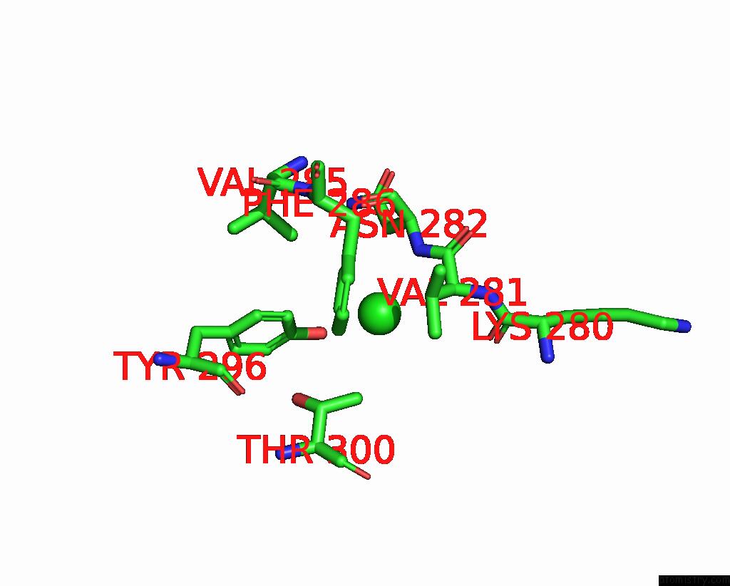
Mono view
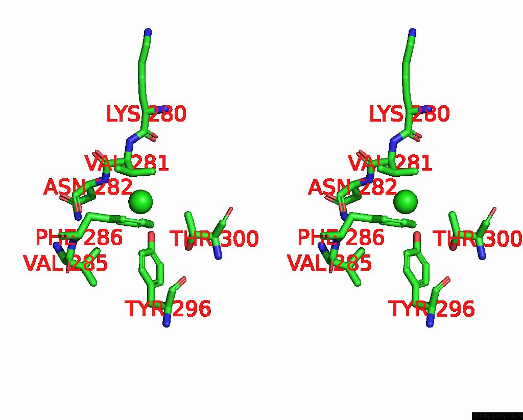
Stereo pair view
Mono view
Stereo pair view
A full contact list of Chlorine with other atoms in the Cl binding
site number 1 of High-Resolution Structure of the Siderophore Periplasmic Binding Protein Ftsb From Streptococcus Pyogenes with Ferrioxamine E Bound (Crystal Form 2) within 5.0Å range:
|
Chlorine binding site 2 out of 2 in 8xfi
Go back to
Chlorine binding site 2 out
of 2 in the High-Resolution Structure of the Siderophore Periplasmic Binding Protein Ftsb From Streptococcus Pyogenes with Ferrioxamine E Bound (Crystal Form 2)
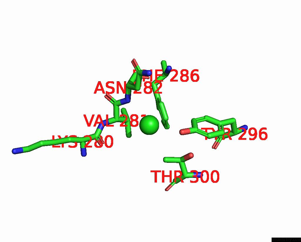
Mono view
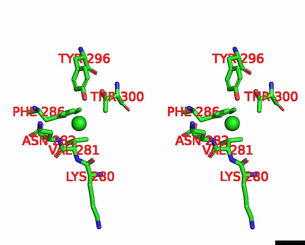
Stereo pair view
Mono view
Stereo pair view
A full contact list of Chlorine with other atoms in the Cl binding
site number 2 of High-Resolution Structure of the Siderophore Periplasmic Binding Protein Ftsb From Streptococcus Pyogenes with Ferrioxamine E Bound (Crystal Form 2) within 5.0Å range:
|
Reference:
J.Fernandez-Perez,
J.M.M.Caaveiro,
A.Senoo,
M.Nakakido,
S.De Vega,
I.Nakagawa,
K.Tsumoto.
Conserved Binding Mechanism For Ligand Promiscuity in the Hydroxamate Siderophore Binding Protein Ftsb From Streptococcus Pyogenes Structure.
ISSN: ISSN 0969-2126
DOI: 10.1016/J.STR.2024.09.018
Page generated: Sun Jul 13 15:41:22 2025
ISSN: ISSN 0969-2126
DOI: 10.1016/J.STR.2024.09.018
Last articles
I in 6PKGI in 6RNS
I in 6RZM
I in 6RQ8
I in 6QE4
I in 6RBC
I in 6QT8
I in 6QFT
I in 6Q4E
I in 6Q4A