Chlorine »
PDB 8xet-8yq4 »
8xuk »
Chlorine in PDB 8xuk: Structure of Beta-1,2-Glucanase From Photobacterium Gaetbulicola (PGSGL3, Ligand-Free)
Protein crystallography data
The structure of Structure of Beta-1,2-Glucanase From Photobacterium Gaetbulicola (PGSGL3, Ligand-Free), PDB code: 8xuk
was solved by
M.Nakajima,
S.Motouchi,
H.Nakai,
with X-Ray Crystallography technique. A brief refinement statistics is given in the table below:
| Resolution Low / High (Å) | 47.21 / 1.20 |
| Space group | P 21 21 21 |
| Cell size a, b, c (Å), α, β, γ (°) | 52.284, 76.595, 94.425, 90, 90, 90 |
| R / Rfree (%) | 19.1 / 20.8 |
Chlorine Binding Sites:
The binding sites of Chlorine atom in the Structure of Beta-1,2-Glucanase From Photobacterium Gaetbulicola (PGSGL3, Ligand-Free)
(pdb code 8xuk). This binding sites where shown within
5.0 Angstroms radius around Chlorine atom.
In total only one binding site of Chlorine was determined in the Structure of Beta-1,2-Glucanase From Photobacterium Gaetbulicola (PGSGL3, Ligand-Free), PDB code: 8xuk:
In total only one binding site of Chlorine was determined in the Structure of Beta-1,2-Glucanase From Photobacterium Gaetbulicola (PGSGL3, Ligand-Free), PDB code: 8xuk:
Chlorine binding site 1 out of 1 in 8xuk
Go back to
Chlorine binding site 1 out
of 1 in the Structure of Beta-1,2-Glucanase From Photobacterium Gaetbulicola (PGSGL3, Ligand-Free)
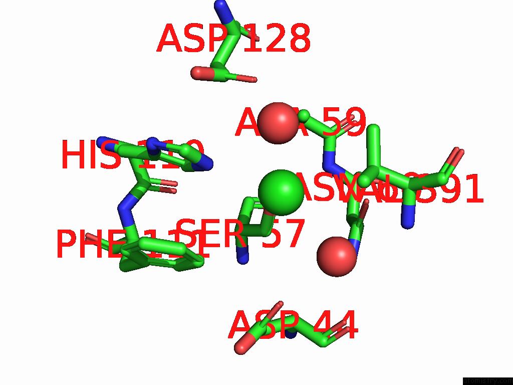
Mono view
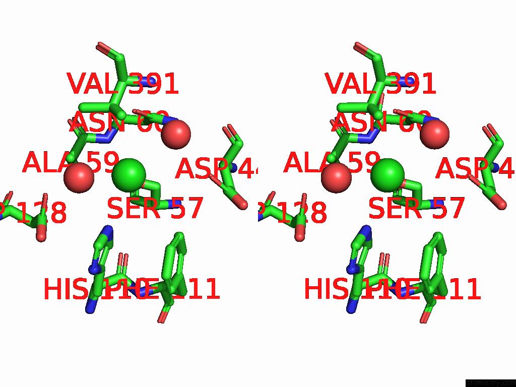
Stereo pair view
Mono view
Stereo pair view
A full contact list of Chlorine with other atoms in the Cl binding
site number 1 of Structure of Beta-1,2-Glucanase From Photobacterium Gaetbulicola (PGSGL3, Ligand-Free) within 5.0Å range:
|
Reference:
M.Nakajima,
N.Tanaka,
S.Motouchi,
K.Kobayashi,
H.Shimizu,
K.Abe,
N.Hosoyamada,
N.Abara,
N.Morimoto,
N.Hiramoto,
R.Nakata,
A.Takashima,
M.Hosoki,
S.Suzuki,
K.Shikano,
T.Fujimaru,
S.Imagawa,
Y.Kawadai,
Z.Wang,
Y.Kitano,
T.Nihira,
H.Nakai,
H.Taguchi.
Beta-1,2-Glucanase Superfamily Identified By Sequential, Functional and Structural Analyses To Be Published.
Page generated: Sun Jul 13 15:44:28 2025
Last articles
Mg in 1IKKMg in 1IK5
Mg in 1IJJ
Mg in 1IJF
Mg in 1IJD
Mg in 1IIR
Mg in 1II9
Mg in 1II6
Mg in 1II0
Mg in 1IHU