Chlorine »
PDB 8yqd-8z8k »
8z26 »
Chlorine in PDB 8z26: Sfx Structure of Cracry 30 Ns After Photoexcitation of the Oxidized Protein
Protein crystallography data
The structure of Sfx Structure of Cracry 30 Ns After Photoexcitation of the Oxidized Protein, PDB code: 8z26
was solved by
M.Maestre-Reyna,
Y.Hosokawa,
P.-H.Wang,
M.Saft,
N.Caramello,
S.Engilberge,
S.Franz-Badur,
E.P.G.Ngura Putu,
M.Nakamura,
W.-J.Wu,
H.-Y.Wu,
C.-C.Lee,
W.-C.Huang,
K.-F.Huang,
Y.-K.Chang,
C.-H.Yang,
W.-T.Lin,
K.-C.Yang,
Y.Ban,
T.Imura,
A.Kazuoka,
E.Tanida,
S.Owada,
Y.Joti,
R.Tanaka,
T.Tanaka,
F.Luo,
K.Tono,
S.Kiontke,
L.Korf,
Y.Umena,
T.Tosha,
Y.Bessho,
E.Nango,
S.Iwata,
A.Royant,
M.-D.Tsai,
J.Yamamoto,
L.-O.Essen,
with X-Ray Crystallography technique. A brief refinement statistics is given in the table below:
| Resolution Low / High (Å) | 30.58 / 2.00 |
| Space group | P 21 21 21 |
| Cell size a, b, c (Å), α, β, γ (°) | 50.84, 65.6, 153.13, 90, 90, 90 |
| R / Rfree (%) | 20.2 / 22.6 |
Chlorine Binding Sites:
The binding sites of Chlorine atom in the Sfx Structure of Cracry 30 Ns After Photoexcitation of the Oxidized Protein
(pdb code 8z26). This binding sites where shown within
5.0 Angstroms radius around Chlorine atom.
In total only one binding site of Chlorine was determined in the Sfx Structure of Cracry 30 Ns After Photoexcitation of the Oxidized Protein, PDB code: 8z26:
In total only one binding site of Chlorine was determined in the Sfx Structure of Cracry 30 Ns After Photoexcitation of the Oxidized Protein, PDB code: 8z26:
Chlorine binding site 1 out of 1 in 8z26
Go back to
Chlorine binding site 1 out
of 1 in the Sfx Structure of Cracry 30 Ns After Photoexcitation of the Oxidized Protein
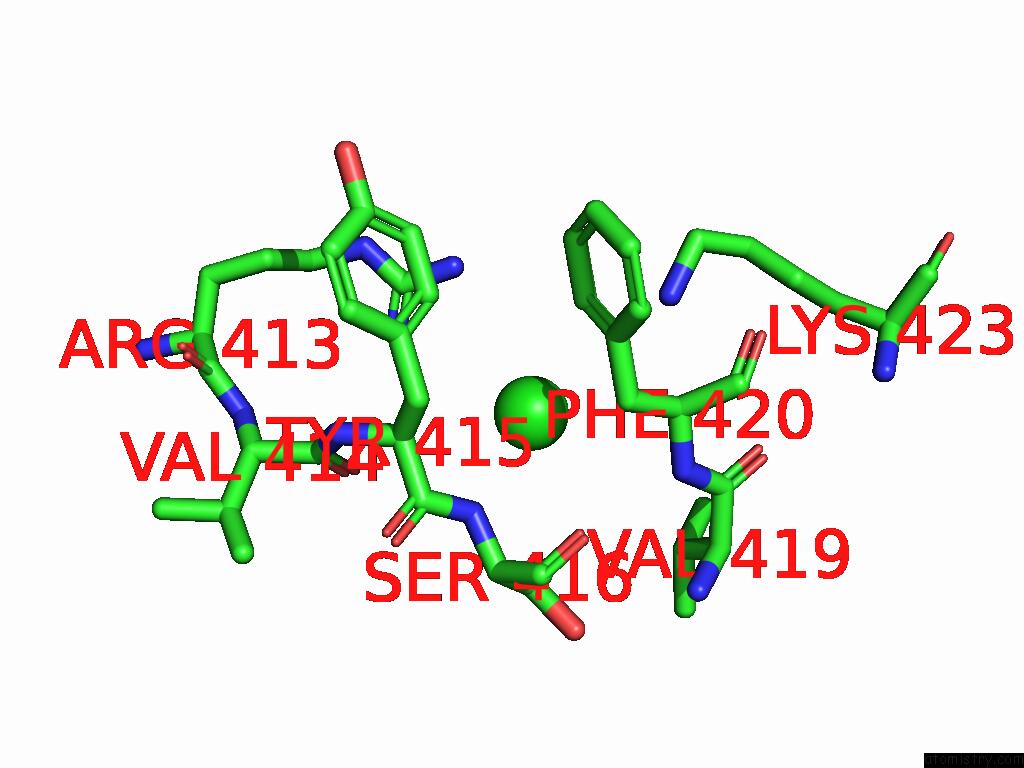
Mono view
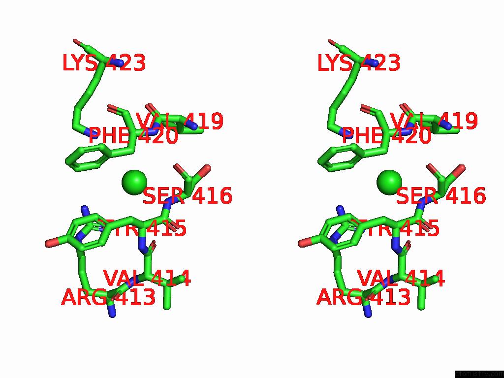
Stereo pair view
Mono view
Stereo pair view
A full contact list of Chlorine with other atoms in the Cl binding
site number 1 of Sfx Structure of Cracry 30 Ns After Photoexcitation of the Oxidized Protein within 5.0Å range:
|
Reference:
M.Maestre-Reyna,
Y.Hosokawa,
P.-H.Wang,
M.Saft,
N.Caramello,
S.Engilberge,
S.Franz-Badur,
E.P.G.Ngura Putu,
M.Nakamura,
W.-J.Wu,
H.-Y.Wu,
C.-C.Lee,
W.-C.Huang,
K.-F.Huang,
Y.-K.Chang,
C.-H.Yang,
W.-T.Lin,
K.-C.Yang,
Y.Ban,
T.Imura,
A.Kazuoka,
E.Tanida,
S.Owada,
Y.Joti,
R.Tanaka,
T.Tanaka,
F.Luo,
K.Tono,
S.Kiontke,
L.Korf,
Y.Umena,
T.Tosha,
Y.Bessho,
E.Nango,
S.Iwata,
A.Royant,
M.-D.Tsai,
J.Yamamoto,
L.-O.Essen.
Capturing Structural Intermediates in An Animal-Like Cryptochrome Photoreceptor By Time-Resolved Crystallography Sci Adv 2025.
ISSN: ESSN 2375-2548
Page generated: Sun Jul 13 15:48:43 2025
ISSN: ESSN 2375-2548
Last articles
I in 4UJ2I in 4UE7
I in 4TVC
I in 4TTU
I in 4TT0
I in 4TTC
I in 4S2V
I in 4S2M
I in 4TKI
I in 4TQD