Chlorine in PDB 9csj: Crystal Structure of Human Glyoxalase Domain-Containing Protein 4 (GLOD4) at 2.33 A Resolution.
Protein crystallography data
The structure of Crystal Structure of Human Glyoxalase Domain-Containing Protein 4 (GLOD4) at 2.33 A Resolution., PDB code: 9csj
was solved by
I.Griswold-Prenner,
Y.Dou,
A.Jennings,
F.Kayser,
with X-Ray Crystallography technique. A brief refinement statistics is given in the table below:
Resolution Low / High (Å)
|
45.34 /
2.33
|
Space group
|
P 41
|
Cell size a, b, c (Å), α, β, γ (°)
|
85.96,
85.96,
135.9,
90,
90,
90
|
R / Rfree (%)
|
22 /
26.9
|
Other elements in 9csj:
The structure of Crystal Structure of Human Glyoxalase Domain-Containing Protein 4 (GLOD4) at 2.33 A Resolution. also contains other interesting chemical elements:
Chlorine Binding Sites:
Pages:
>>> Page 1 <<<
Page 2, Binding sites: 11 -
12;
Binding sites:
The binding sites of Chlorine atom in the Crystal Structure of Human Glyoxalase Domain-Containing Protein 4 (GLOD4) at 2.33 A Resolution.
(pdb code 9csj). This binding sites where shown within
5.0 Angstroms radius around Chlorine atom.
In total 12 binding sites of Chlorine where determined in the
Crystal Structure of Human Glyoxalase Domain-Containing Protein 4 (GLOD4) at 2.33 A Resolution., PDB code: 9csj:
Jump to Chlorine binding site number:
1;
2;
3;
4;
5;
6;
7;
8;
9;
10;
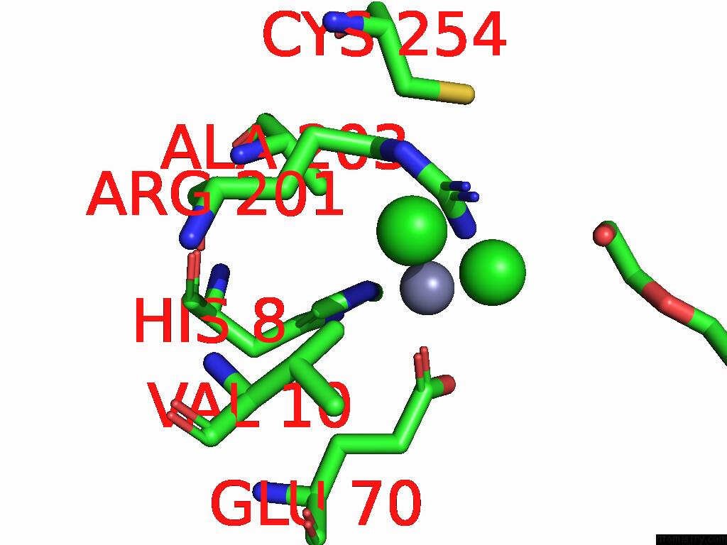
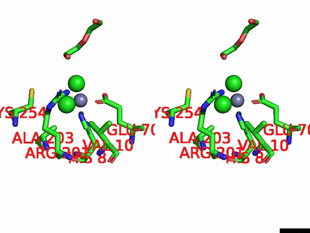
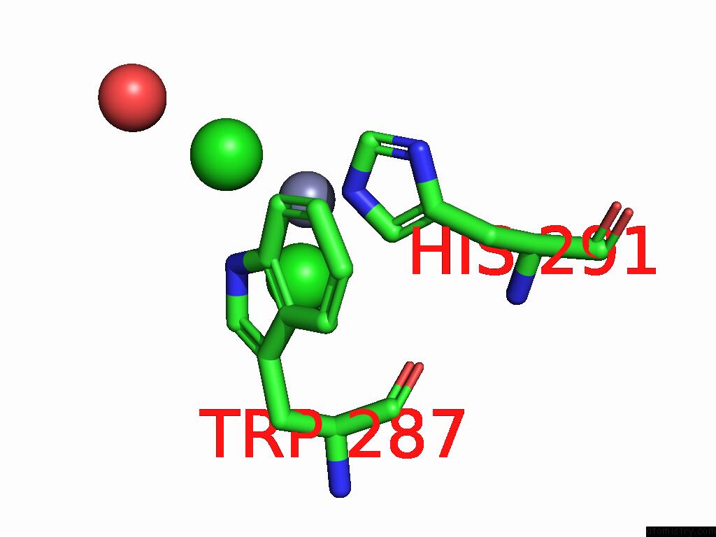
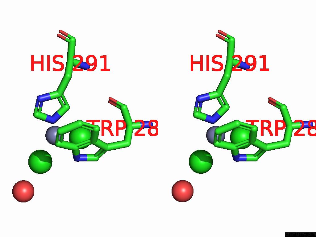
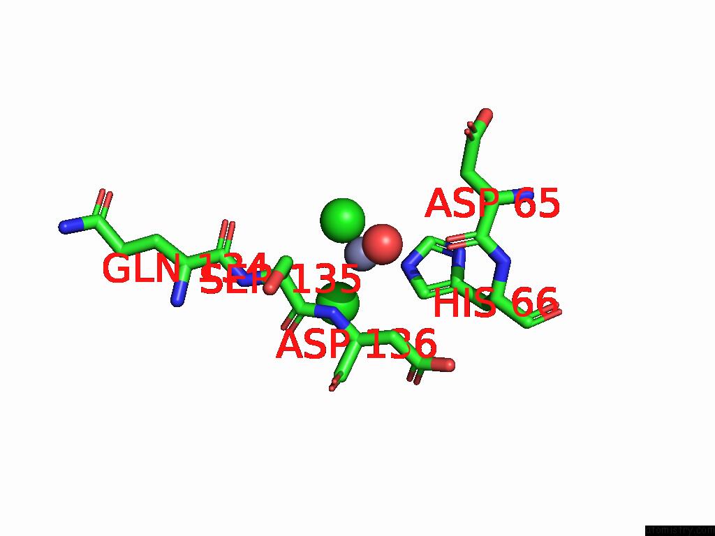
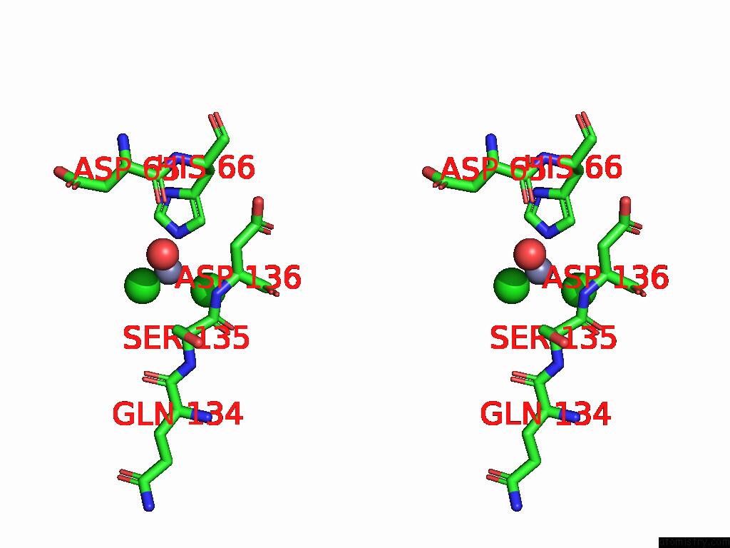
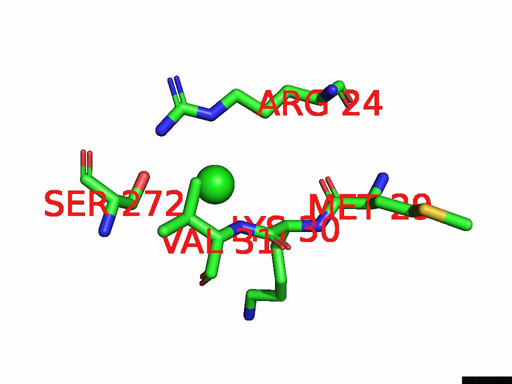
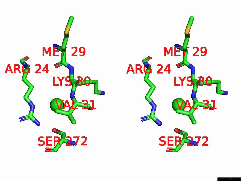
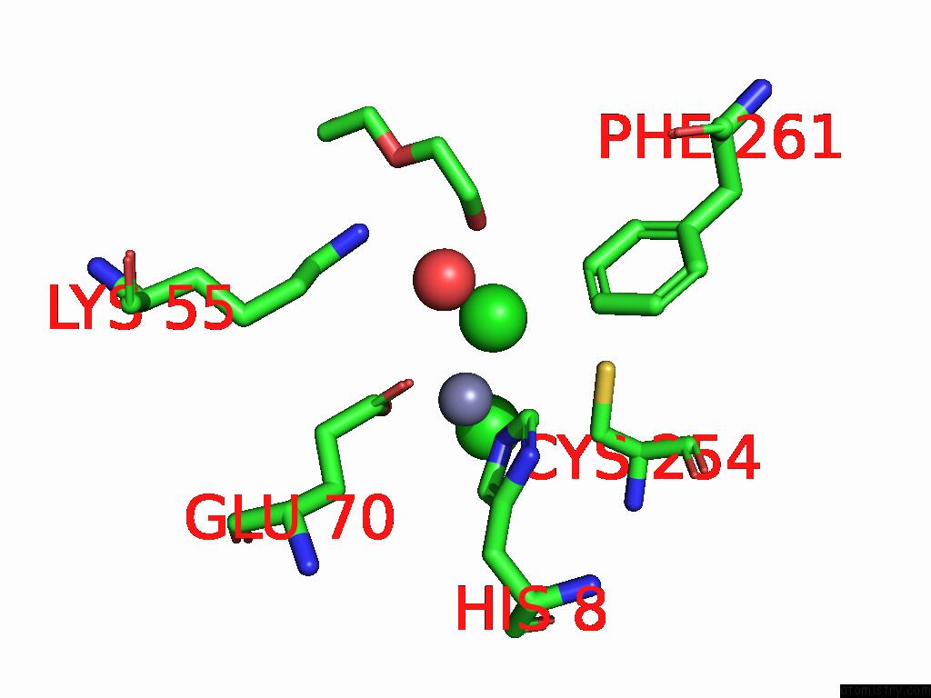
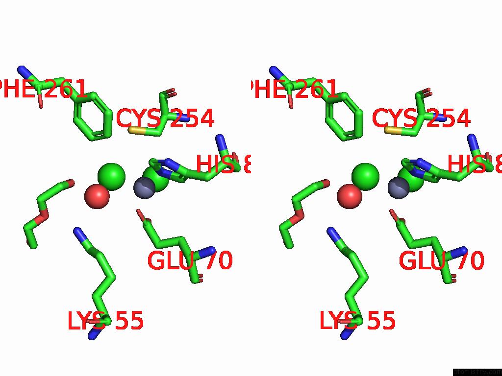
Chlorine binding site 1 out
of 12 in 9csj
Go back to
Chlorine Binding Sites List in 9csj
Chlorine binding site 1 out
of 12 in the Crystal Structure of Human Glyoxalase Domain-Containing Protein 4 (GLOD4) at 2.33 A Resolution.
 Mono view
 Stereo pair view
|
A full contact list of Chlorine with other atoms in the Cl binding
site number 1 of Crystal Structure of Human Glyoxalase Domain-Containing Protein 4 (GLOD4) at 2.33 A Resolution. within 5.0Å range:
probe
|
atom
|
residue
|
distance (Å)
|
B
|
Occ
|
A:Cl305
b:36.9
occ:1.00
|
ZN
|
A:ZN302
|
2.1
|
35.7
|
1.0
|
OE1
|
A:GLU70
|
3.1
|
34.7
|
1.0
|
NE2
|
A:HIS8
|
3.5
|
30.8
|
1.0
|
CD
|
A:ARG201
|
3.6
|
34.5
|
1.0
|
CG2
|
A:VAL10
|
3.6
|
28.7
|
1.0
|
CL
|
A:CL310
|
3.6
|
46.4
|
1.0
|
CB
|
A:CYS254
|
3.6
|
33.2
|
1.0
|
NH2
|
A:ARG201
|
3.7
|
39.2
|
1.0
|
CB
|
A:ALA203
|
3.9
|
28.9
|
1.0
|
SG
|
A:CYS254
|
4.0
|
33.4
|
1.0
|
CD2
|
A:HIS8
|
4.1
|
31.1
|
1.0
|
CD
|
A:GLU70
|
4.2
|
35.6
|
1.0
|
CG
|
A:ARG201
|
4.4
|
32.9
|
1.0
|
NE
|
A:ARG201
|
4.5
|
36.3
|
1.0
|
CZ
|
A:ARG201
|
4.6
|
38.4
|
1.0
|
CE1
|
A:HIS8
|
4.6
|
32.2
|
1.0
|
OE2
|
A:GLU70
|
4.6
|
41.9
|
1.0
|
CA
|
A:ALA203
|
4.8
|
28.8
|
1.0
|
O4
|
A:PEG312
|
4.8
|
57.2
|
1.0
|
CB
|
A:ARG201
|
4.8
|
31.1
|
1.0
|
|
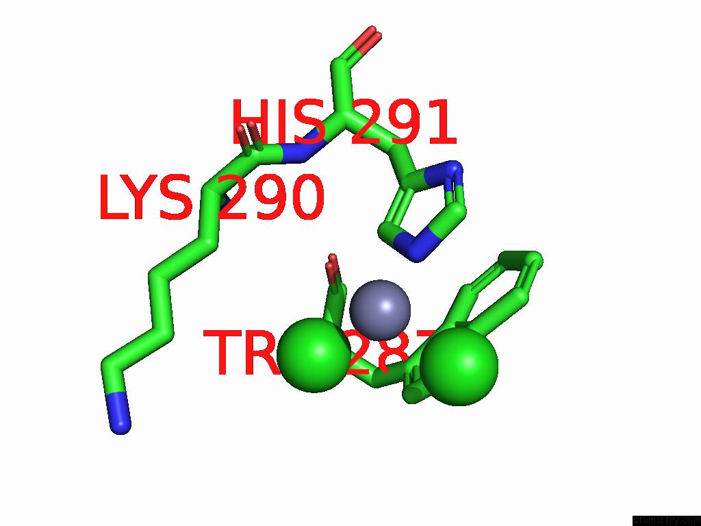
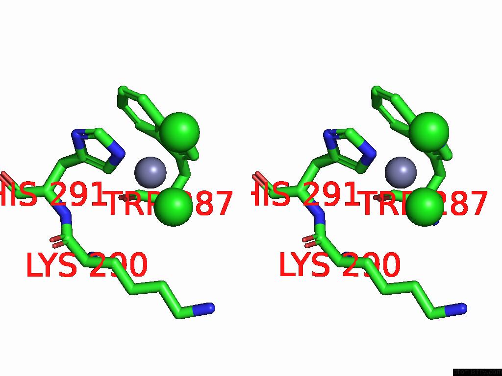
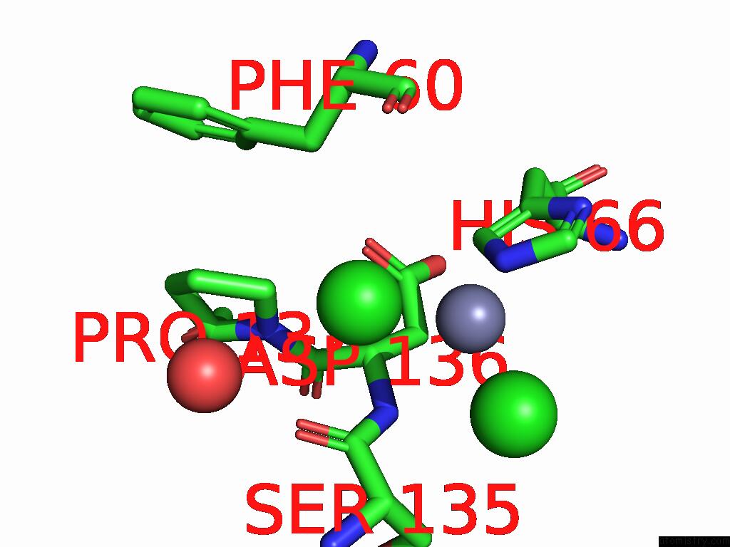
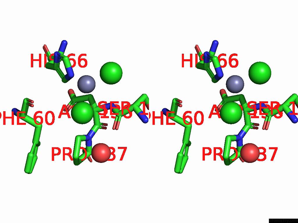
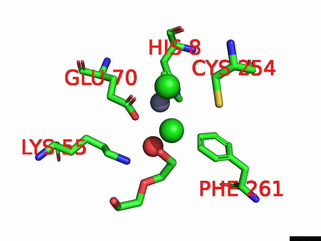
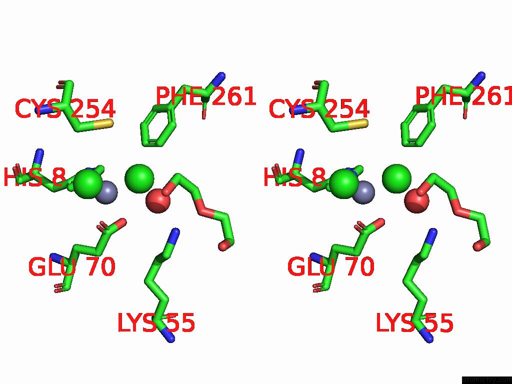
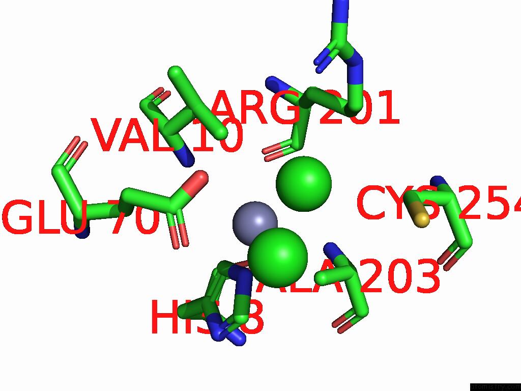
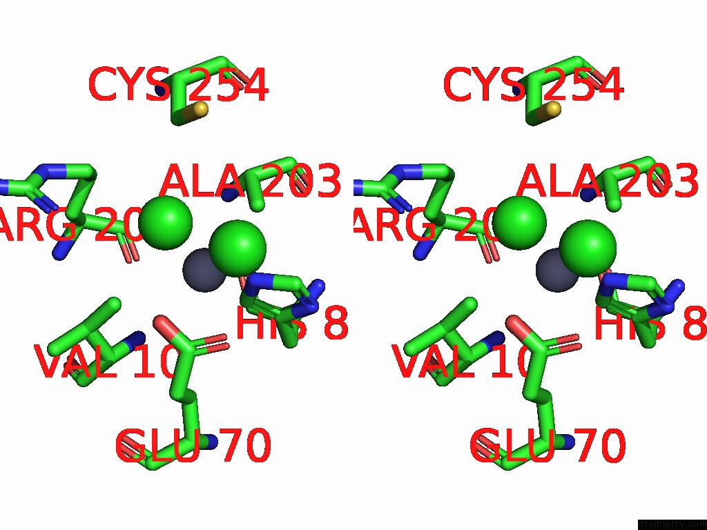
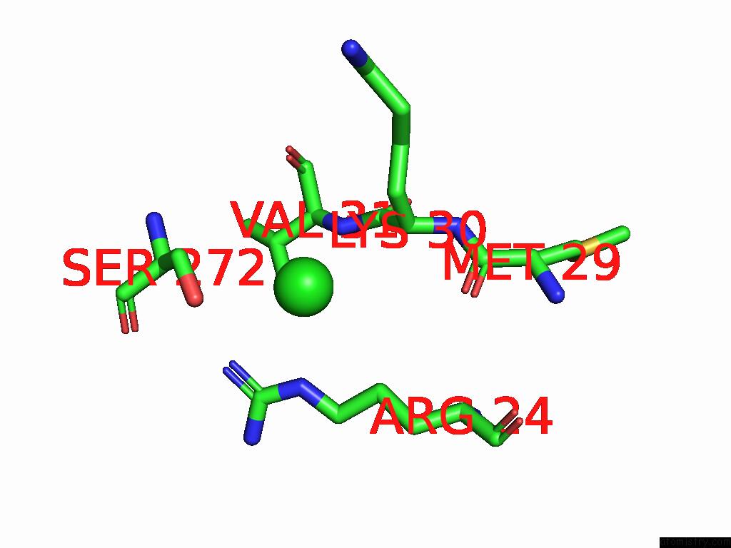
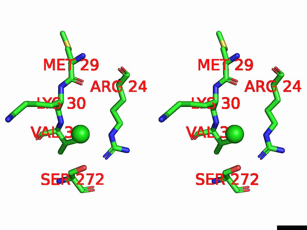
Chlorine binding site 2 out
of 12 in 9csj
Go back to
Chlorine Binding Sites List in 9csj
Chlorine binding site 2 out
of 12 in the Crystal Structure of Human Glyoxalase Domain-Containing Protein 4 (GLOD4) at 2.33 A Resolution.
 Mono view
 Stereo pair view
|
A full contact list of Chlorine with other atoms in the Cl binding
site number 2 of Crystal Structure of Human Glyoxalase Domain-Containing Protein 4 (GLOD4) at 2.33 A Resolution. within 5.0Å range:
probe
|
atom
|
residue
|
distance (Å)
|
B
|
Occ
|
A:Cl306
b:32.0
occ:1.00
|
ZN
|
A:ZN304
|
2.1
|
29.3
|
1.0
|
NE2
|
A:HIS291
|
3.4
|
31.6
|
1.0
|
CD1
|
A:TRP287
|
3.5
|
32.0
|
1.0
|
CD2
|
A:HIS291
|
3.6
|
32.3
|
1.0
|
NE1
|
A:TRP287
|
3.7
|
33.1
|
1.0
|
CL
|
A:CL307
|
3.8
|
33.9
|
1.0
|
CD
|
A:LYS290
|
4.0
|
39.5
|
1.0
|
CB
|
A:LYS290
|
4.1
|
34.5
|
1.0
|
CG
|
A:TRP287
|
4.3
|
31.0
|
1.0
|
CE2
|
A:TRP287
|
4.5
|
32.7
|
1.0
|
O
|
A:TRP287
|
4.6
|
28.2
|
1.0
|
CA
|
A:TRP287
|
4.6
|
30.9
|
1.0
|
CE1
|
A:HIS291
|
4.7
|
31.7
|
1.0
|
CG
|
A:LYS290
|
4.7
|
37.0
|
1.0
|
CD2
|
A:TRP287
|
4.9
|
30.8
|
1.0
|
CG
|
A:HIS291
|
5.0
|
31.2
|
1.0
|
CB
|
A:TRP287
|
5.0
|
31.0
|
1.0
|
|
Chlorine binding site 3 out
of 12 in 9csj
Go back to
Chlorine Binding Sites List in 9csj
Chlorine binding site 3 out
of 12 in the Crystal Structure of Human Glyoxalase Domain-Containing Protein 4 (GLOD4) at 2.33 A Resolution.
 Mono view
 Stereo pair view
|
A full contact list of Chlorine with other atoms in the Cl binding
site number 3 of Crystal Structure of Human Glyoxalase Domain-Containing Protein 4 (GLOD4) at 2.33 A Resolution. within 5.0Å range:
probe
|
atom
|
residue
|
distance (Å)
|
B
|
Occ
|
A:Cl307
b:33.9
occ:1.00
|
ZN
|
A:ZN304
|
2.3
|
29.3
|
1.0
|
NE1
|
A:TRP287
|
3.3
|
33.1
|
1.0
|
NE2
|
A:HIS291
|
3.4
|
31.6
|
1.0
|
O
|
A:HOH584
|
3.4
|
35.6
|
1.0
|
CL
|
A:CL306
|
3.8
|
32.0
|
1.0
|
CE1
|
A:HIS291
|
3.8
|
31.7
|
1.0
|
CE2
|
A:TRP287
|
4.1
|
32.7
|
1.0
|
CZ2
|
A:TRP287
|
4.3
|
33.0
|
1.0
|
CD1
|
A:TRP287
|
4.3
|
32.0
|
1.0
|
CD2
|
A:HIS291
|
4.6
|
32.3
|
1.0
|
|
Chlorine binding site 4 out
of 12 in 9csj
Go back to
Chlorine Binding Sites List in 9csj
Chlorine binding site 4 out
of 12 in the Crystal Structure of Human Glyoxalase Domain-Containing Protein 4 (GLOD4) at 2.33 A Resolution.
 Mono view
 Stereo pair view
|
A full contact list of Chlorine with other atoms in the Cl binding
site number 4 of Crystal Structure of Human Glyoxalase Domain-Containing Protein 4 (GLOD4) at 2.33 A Resolution. within 5.0Å range:
probe
|
atom
|
residue
|
distance (Å)
|
B
|
Occ
|
A:Cl308
b:36.1
occ:1.00
|
ZN
|
A:ZN303
|
2.3
|
30.3
|
1.0
|
NE2
|
A:HIS66
|
3.3
|
31.2
|
1.0
|
O
|
A:HOH515
|
3.5
|
34.0
|
1.0
|
O
|
A:SER135
|
3.5
|
35.6
|
1.0
|
CD
|
A:PRO137
|
3.6
|
29.7
|
1.0
|
CD2
|
A:HIS66
|
3.7
|
30.8
|
1.0
|
C
|
A:SER135
|
3.8
|
35.2
|
1.0
|
CL
|
A:CL309
|
3.9
|
32.3
|
1.0
|
CA
|
A:ASP136
|
3.9
|
30.5
|
1.0
|
CB
|
A:PHE60
|
4.0
|
29.6
|
1.0
|
N
|
A:ASP136
|
4.0
|
32.5
|
1.0
|
O
|
A:PHE60
|
4.1
|
28.6
|
1.0
|
CG
|
A:PRO137
|
4.5
|
30.9
|
1.0
|
C
|
A:PHE60
|
4.5
|
27.7
|
1.0
|
CE1
|
A:HIS66
|
4.5
|
31.6
|
1.0
|
CA
|
A:SER135
|
4.5
|
37.0
|
1.0
|
N
|
A:PRO137
|
4.6
|
29.8
|
1.0
|
C
|
A:ASP136
|
4.7
|
30.2
|
1.0
|
CA
|
A:PHE60
|
4.9
|
28.1
|
1.0
|
CB
|
A:ASP136
|
4.9
|
30.4
|
1.0
|
CG
|
A:PHE60
|
5.0
|
31.1
|
1.0
|
|
Chlorine binding site 5 out
of 12 in 9csj
Go back to
Chlorine Binding Sites List in 9csj
Chlorine binding site 5 out
of 12 in the Crystal Structure of Human Glyoxalase Domain-Containing Protein 4 (GLOD4) at 2.33 A Resolution.
 Mono view
 Stereo pair view
|
A full contact list of Chlorine with other atoms in the Cl binding
site number 5 of Crystal Structure of Human Glyoxalase Domain-Containing Protein 4 (GLOD4) at 2.33 A Resolution. within 5.0Å range:
probe
|
atom
|
residue
|
distance (Å)
|
B
|
Occ
|
A:Cl309
b:32.3
occ:1.00
|
ZN
|
A:ZN303
|
2.1
|
30.3
|
1.0
|
O
|
A:HOH441
|
3.2
|
31.2
|
1.0
|
NE2
|
A:HIS66
|
3.4
|
31.2
|
1.0
|
CA
|
A:SER135
|
3.7
|
37.0
|
1.0
|
CE1
|
A:HIS66
|
3.8
|
31.6
|
1.0
|
CL
|
A:CL308
|
3.9
|
36.1
|
1.0
|
C
|
A:SER135
|
4.1
|
35.2
|
1.0
|
N
|
A:ASP136
|
4.1
|
32.5
|
1.0
|
CB
|
A:SER135
|
4.3
|
38.1
|
1.0
|
CD2
|
A:HIS66
|
4.4
|
30.8
|
1.0
|
O
|
A:GLN134
|
4.6
|
48.6
|
1.0
|
N
|
A:SER135
|
4.7
|
38.5
|
1.0
|
O
|
A:ASP65
|
4.8
|
30.8
|
1.0
|
O
|
A:SER135
|
4.9
|
35.6
|
1.0
|
ND1
|
A:HIS66
|
4.9
|
29.4
|
1.0
|
CA
|
A:ASP136
|
5.0
|
30.5
|
1.0
|
|
Chlorine binding site 6 out
of 12 in 9csj
Go back to
Chlorine Binding Sites List in 9csj
Chlorine binding site 6 out
of 12 in the Crystal Structure of Human Glyoxalase Domain-Containing Protein 4 (GLOD4) at 2.33 A Resolution.
 Mono view
 Stereo pair view
|
A full contact list of Chlorine with other atoms in the Cl binding
site number 6 of Crystal Structure of Human Glyoxalase Domain-Containing Protein 4 (GLOD4) at 2.33 A Resolution. within 5.0Å range:
probe
|
atom
|
residue
|
distance (Å)
|
B
|
Occ
|
A:Cl310
b:46.4
occ:1.00
|
ZN
|
A:ZN302
|
2.2
|
35.7
|
1.0
|
O
|
A:HOH459
|
2.9
|
36.9
|
1.0
|
OE2
|
A:GLU70
|
3.3
|
41.9
|
1.0
|
C4
|
A:PEG312
|
3.3
|
55.7
|
1.0
|
NE2
|
A:HIS8
|
3.4
|
30.8
|
1.0
|
CL
|
A:CL305
|
3.6
|
36.9
|
1.0
|
CE1
|
A:HIS8
|
3.6
|
32.2
|
1.0
|
OE1
|
A:GLU70
|
3.6
|
34.7
|
1.0
|
C3
|
A:PEG312
|
3.7
|
57.0
|
1.0
|
O4
|
A:PEG312
|
3.8
|
57.2
|
1.0
|
CD
|
A:GLU70
|
3.9
|
35.6
|
1.0
|
NZ
|
A:LYS55
|
3.9
|
41.8
|
1.0
|
CE1
|
A:PHE261
|
4.3
|
28.7
|
1.0
|
CD2
|
A:HIS8
|
4.7
|
31.1
|
1.0
|
O2
|
A:PEG312
|
4.7
|
57.7
|
1.0
|
ND1
|
A:HIS8
|
4.9
|
29.9
|
1.0
|
CE
|
A:LYS55
|
4.9
|
39.8
|
1.0
|
SG
|
A:CYS254
|
4.9
|
33.4
|
1.0
|
|
Chlorine binding site 7 out
of 12 in 9csj
Go back to
Chlorine Binding Sites List in 9csj
Chlorine binding site 7 out
of 12 in the Crystal Structure of Human Glyoxalase Domain-Containing Protein 4 (GLOD4) at 2.33 A Resolution.
 Mono view
 Stereo pair view
|
A full contact list of Chlorine with other atoms in the Cl binding
site number 7 of Crystal Structure of Human Glyoxalase Domain-Containing Protein 4 (GLOD4) at 2.33 A Resolution. within 5.0Å range:
probe
|
atom
|
residue
|
distance (Å)
|
B
|
Occ
|
A:Cl311
b:40.7
occ:1.00
|
N
|
A:VAL31
|
3.1
|
31.8
|
1.0
|
OG
|
A:SER272
|
3.3
|
43.2
|
1.0
|
NE
|
A:ARG24
|
3.3
|
36.9
|
1.0
|
NH2
|
A:ARG24
|
3.6
|
39.9
|
1.0
|
CA
|
A:LYS30
|
3.8
|
34.6
|
1.0
|
CG2
|
A:VAL31
|
3.8
|
33.6
|
1.0
|
CB
|
A:VAL31
|
3.8
|
32.1
|
1.0
|
CB
|
A:SER272
|
3.8
|
39.8
|
1.0
|
CZ
|
A:ARG24
|
3.9
|
38.5
|
1.0
|
C
|
A:LYS30
|
3.9
|
33.0
|
1.0
|
CB
|
A:LYS30
|
4.0
|
37.6
|
1.0
|
CG
|
A:ARG24
|
4.0
|
34.5
|
1.0
|
CA
|
A:VAL31
|
4.0
|
31.6
|
1.0
|
CD
|
A:ARG24
|
4.2
|
36.6
|
1.0
|
CA
|
A:SER272
|
4.7
|
39.5
|
1.0
|
O
|
A:VAL31
|
4.7
|
29.9
|
1.0
|
C
|
A:VAL31
|
4.9
|
30.3
|
1.0
|
O
|
A:MET29
|
5.0
|
30.6
|
1.0
|
|
Chlorine binding site 8 out
of 12 in 9csj
Go back to
Chlorine Binding Sites List in 9csj
Chlorine binding site 8 out
of 12 in the Crystal Structure of Human Glyoxalase Domain-Containing Protein 4 (GLOD4) at 2.33 A Resolution.
 Mono view
 Stereo pair view
|
A full contact list of Chlorine with other atoms in the Cl binding
site number 8 of Crystal Structure of Human Glyoxalase Domain-Containing Protein 4 (GLOD4) at 2.33 A Resolution. within 5.0Å range:
probe
|
atom
|
residue
|
distance (Å)
|
B
|
Occ
|
B:Cl305
b:35.5
occ:1.00
|
ZN
|
B:ZN304
|
2.1
|
35.6
|
1.0
|
OE2
|
B:GLU70
|
3.4
|
37.9
|
1.0
|
NE2
|
B:HIS8
|
3.5
|
33.0
|
1.0
|
CD
|
B:ARG201
|
3.6
|
38.4
|
1.0
|
CB
|
B:CYS254
|
3.6
|
32.0
|
1.0
|
CL
|
B:CL306
|
3.6
|
48.8
|
1.0
|
CG2
|
B:VAL10
|
3.7
|
29.1
|
1.0
|
NH1
|
B:ARG201
|
3.7
|
43.2
|
1.0
|
CB
|
B:ALA203
|
3.9
|
29.7
|
1.0
|
SG
|
B:CYS254
|
3.9
|
33.1
|
1.0
|
CD2
|
B:HIS8
|
4.1
|
33.0
|
1.0
|
CD
|
B:GLU70
|
4.3
|
36.4
|
1.0
|
CG
|
B:ARG201
|
4.4
|
35.6
|
1.0
|
OE1
|
B:GLU70
|
4.5
|
38.7
|
1.0
|
NE
|
B:ARG201
|
4.5
|
41.2
|
1.0
|
CZ
|
B:ARG201
|
4.6
|
43.3
|
1.0
|
CE1
|
B:HIS8
|
4.7
|
33.1
|
1.0
|
CA
|
B:ALA203
|
4.7
|
29.3
|
1.0
|
CB
|
B:ARG201
|
4.9
|
33.9
|
1.0
|
|
Chlorine binding site 9 out
of 12 in 9csj
Go back to
Chlorine Binding Sites List in 9csj
Chlorine binding site 9 out
of 12 in the Crystal Structure of Human Glyoxalase Domain-Containing Protein 4 (GLOD4) at 2.33 A Resolution.
 Mono view
 Stereo pair view
|
A full contact list of Chlorine with other atoms in the Cl binding
site number 9 of Crystal Structure of Human Glyoxalase Domain-Containing Protein 4 (GLOD4) at 2.33 A Resolution. within 5.0Å range:
probe
|
atom
|
residue
|
distance (Å)
|
B
|
Occ
|
B:Cl306
b:48.8
occ:1.00
|
ZN
|
B:ZN304
|
2.3
|
35.6
|
1.0
|
O
|
B:HOH516
|
2.9
|
39.3
|
1.0
|
OE1
|
B:GLU70
|
3.2
|
38.7
|
1.0
|
NE2
|
B:HIS8
|
3.4
|
33.0
|
1.0
|
O4
|
B:PEG309
|
3.5
|
59.7
|
1.0
|
C4
|
B:PEG309
|
3.5
|
60.1
|
1.0
|
CL
|
B:CL305
|
3.6
|
35.5
|
1.0
|
CE1
|
B:HIS8
|
3.6
|
33.1
|
1.0
|
OE2
|
B:GLU70
|
3.7
|
37.9
|
1.0
|
CD
|
B:GLU70
|
3.9
|
36.4
|
1.0
|
NZ
|
B:LYS55
|
4.1
|
38.8
|
1.0
|
CE1
|
B:PHE261
|
4.4
|
30.3
|
1.0
|
CD2
|
B:HIS8
|
4.7
|
33.0
|
1.0
|
CE
|
B:LYS55
|
4.9
|
36.0
|
1.0
|
C3
|
B:PEG309
|
4.9
|
60.9
|
1.0
|
ND1
|
B:HIS8
|
4.9
|
31.7
|
1.0
|
SG
|
B:CYS254
|
4.9
|
33.1
|
1.0
|
|
Chlorine binding site 10 out
of 12 in 9csj
Go back to
Chlorine Binding Sites List in 9csj
Chlorine binding site 10 out
of 12 in the Crystal Structure of Human Glyoxalase Domain-Containing Protein 4 (GLOD4) at 2.33 A Resolution.
 Mono view
 Stereo pair view
|
A full contact list of Chlorine with other atoms in the Cl binding
site number 10 of Crystal Structure of Human Glyoxalase Domain-Containing Protein 4 (GLOD4) at 2.33 A Resolution. within 5.0Å range:
probe
|
atom
|
residue
|
distance (Å)
|
B
|
Occ
|
B:Cl307
b:41.4
occ:1.00
|
N
|
B:VAL31
|
3.1
|
33.3
|
1.0
|
OG
|
B:SER272
|
3.2
|
45.2
|
1.0
|
NE
|
B:ARG24
|
3.3
|
35.3
|
1.0
|
NH1
|
B:ARG24
|
3.7
|
40.3
|
1.0
|
CA
|
B:LYS30
|
3.7
|
36.6
|
1.0
|
CG2
|
B:VAL31
|
3.8
|
36.8
|
1.0
|
CB
|
B:SER272
|
3.8
|
39.0
|
1.0
|
CB
|
B:VAL31
|
3.9
|
34.9
|
1.0
|
C
|
B:LYS30
|
3.9
|
34.6
|
1.0
|
CB
|
B:LYS30
|
3.9
|
37.3
|
1.0
|
CZ
|
B:ARG24
|
3.9
|
38.0
|
1.0
|
CG
|
B:ARG24
|
4.0
|
34.5
|
1.0
|
CA
|
B:VAL31
|
4.1
|
33.0
|
1.0
|
CD
|
B:ARG24
|
4.2
|
36.2
|
1.0
|
O
|
B:VAL31
|
4.7
|
30.2
|
1.0
|
CA
|
B:SER272
|
4.7
|
38.1
|
1.0
|
C
|
B:VAL31
|
4.9
|
31.1
|
1.0
|
O
|
B:MET29
|
4.9
|
34.5
|
1.0
|
|
Reference:
I.Griswold-Prenner,
F.Kayser.
Crystal Structure of Apo-Glyoxalase Domain-Containing Protein 4 (Apo-GLOD4) at 2.33 A Resolution. To Be Published.
Page generated: Sat Aug 23 00:39:20 2025
|