Chlorine »
PDB 9coz-9dpt »
9da9 »
Chlorine in PDB 9da9: Crystal Structure of GLUN1/GLUN2A Agonist-Binding Domains in Complex with 7CKA and Glutamate
Protein crystallography data
The structure of Crystal Structure of GLUN1/GLUN2A Agonist-Binding Domains in Complex with 7CKA and Glutamate, PDB code: 9da9
was solved by
J.Bosco,
C.K.Yates-Hansen,
L.J.Mcclelland,
E.Voronina,
K.B.Hansen,
with X-Ray Crystallography technique. A brief refinement statistics is given in the table below:
| Resolution Low / High (Å) | 33.97 / 2.05 |
| Space group | P 21 21 21 |
| Cell size a, b, c (Å), α, β, γ (°) | 54.244, 87.338, 135.896, 90, 90, 90 |
| R / Rfree (%) | 16.8 / 22.8 |
Chlorine Binding Sites:
The binding sites of Chlorine atom in the Crystal Structure of GLUN1/GLUN2A Agonist-Binding Domains in Complex with 7CKA and Glutamate
(pdb code 9da9). This binding sites where shown within
5.0 Angstroms radius around Chlorine atom.
In total only one binding site of Chlorine was determined in the Crystal Structure of GLUN1/GLUN2A Agonist-Binding Domains in Complex with 7CKA and Glutamate, PDB code: 9da9:
In total only one binding site of Chlorine was determined in the Crystal Structure of GLUN1/GLUN2A Agonist-Binding Domains in Complex with 7CKA and Glutamate, PDB code: 9da9:
Chlorine binding site 1 out of 1 in 9da9
Go back to
Chlorine binding site 1 out
of 1 in the Crystal Structure of GLUN1/GLUN2A Agonist-Binding Domains in Complex with 7CKA and Glutamate
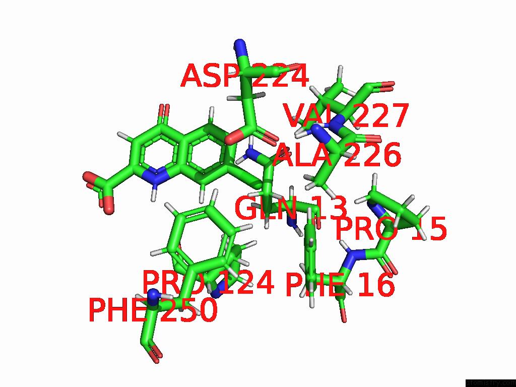
Mono view
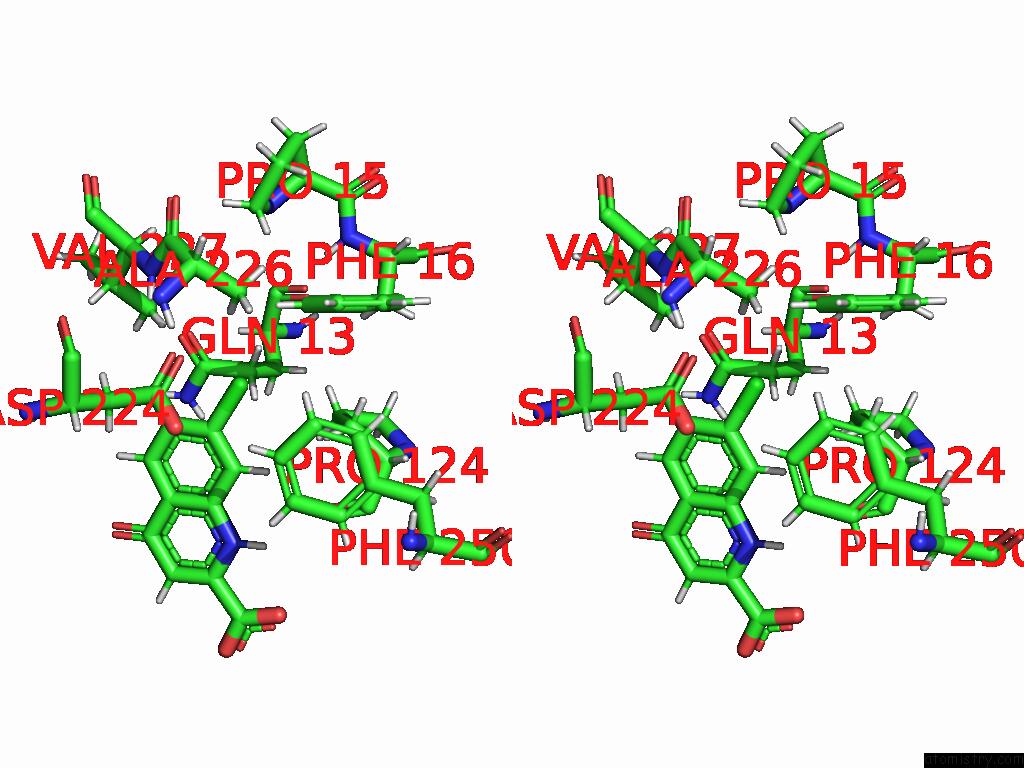
Stereo pair view
Mono view
Stereo pair view
A full contact list of Chlorine with other atoms in the Cl binding
site number 1 of Crystal Structure of GLUN1/GLUN2A Agonist-Binding Domains in Complex with 7CKA and Glutamate within 5.0Å range:
|
Reference:
J.Bosco,
C.K.Yates-Hansen,
L.J.Mcclelland,
E.Voronina,
K.B.Hansen.
Crystal Structure of GLUN1/GLUN2A Agonist-Binding Domains in Complex with 7CKA and Glutamate Nature 2025.
ISSN: ESSN 1476-4687
DOI: 10.1038/S41598-025-91954-5
Page generated: Sun Jul 13 16:14:36 2025
ISSN: ESSN 1476-4687
DOI: 10.1038/S41598-025-91954-5
Last articles
Fe in 7MJXFe in 7MJW
Fe in 7MK8
Fe in 7MJZ
Fe in 7MK4
Fe in 7MJY
Fe in 7MI4
Fe in 7MJV
Fe in 7MI5
Fe in 7MID