Chlorine »
PDB 9ger-9hh3 »
9hgo »
Chlorine in PDB 9hgo: Crystal Structure of the Coxiella Burnetii E110Q Mutant 2- Methylisocitrate Lyase
Enzymatic activity of Crystal Structure of the Coxiella Burnetii E110Q Mutant 2- Methylisocitrate Lyase
All present enzymatic activity of Crystal Structure of the Coxiella Burnetii E110Q Mutant 2- Methylisocitrate Lyase:
4.1.3.30;
4.1.3.30;
Protein crystallography data
The structure of Crystal Structure of the Coxiella Burnetii E110Q Mutant 2- Methylisocitrate Lyase, PDB code: 9hgo
was solved by
W.Stuart,
M.Isupov,
N.J.Harmer,
with X-Ray Crystallography technique. A brief refinement statistics is given in the table below:
| Resolution Low / High (Å) | 61.48 / 1.82 |
| Space group | P 31 2 1 |
| Cell size a, b, c (Å), α, β, γ (°) | 74.18, 74.18, 184.45, 90, 90, 120 |
| R / Rfree (%) | 23.2 / 29.9 |
Chlorine Binding Sites:
The binding sites of Chlorine atom in the Crystal Structure of the Coxiella Burnetii E110Q Mutant 2- Methylisocitrate Lyase
(pdb code 9hgo). This binding sites where shown within
5.0 Angstroms radius around Chlorine atom.
In total 2 binding sites of Chlorine where determined in the Crystal Structure of the Coxiella Burnetii E110Q Mutant 2- Methylisocitrate Lyase, PDB code: 9hgo:
Jump to Chlorine binding site number: 1; 2;
In total 2 binding sites of Chlorine where determined in the Crystal Structure of the Coxiella Burnetii E110Q Mutant 2- Methylisocitrate Lyase, PDB code: 9hgo:
Jump to Chlorine binding site number: 1; 2;
Chlorine binding site 1 out of 2 in 9hgo
Go back to
Chlorine binding site 1 out
of 2 in the Crystal Structure of the Coxiella Burnetii E110Q Mutant 2- Methylisocitrate Lyase
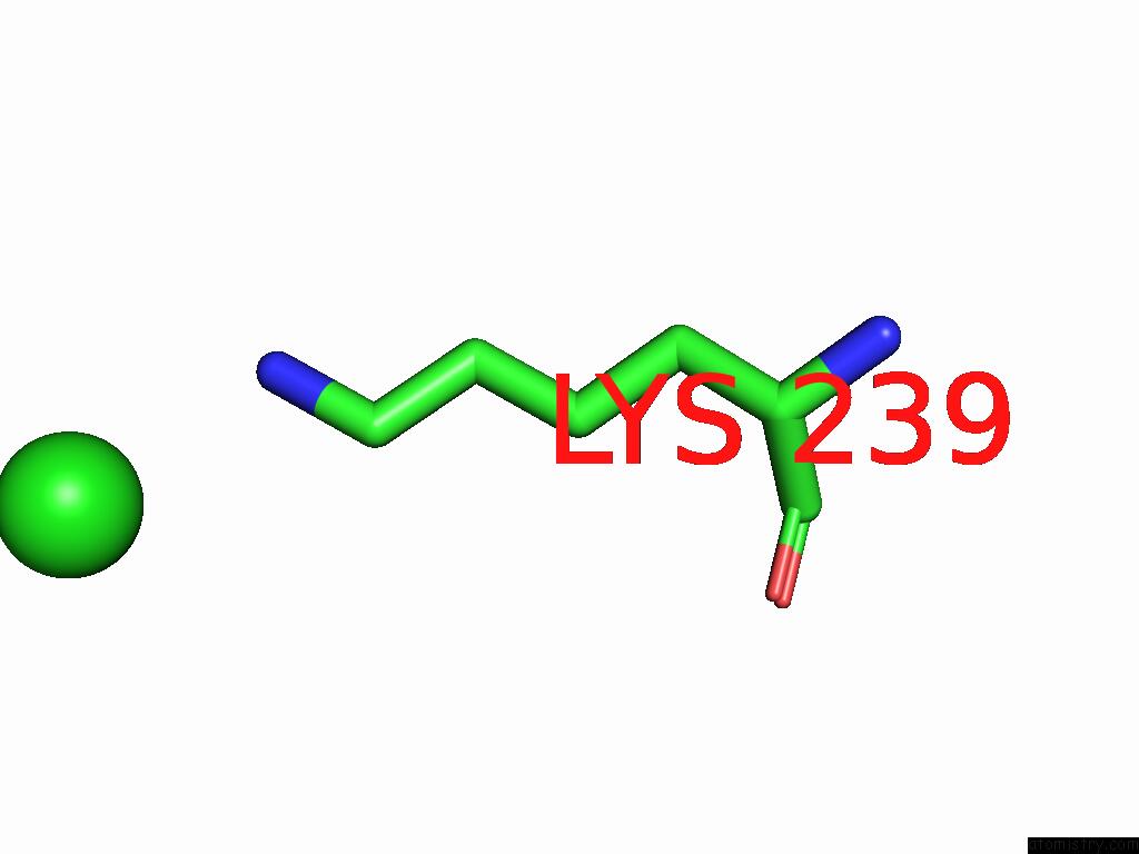
Mono view
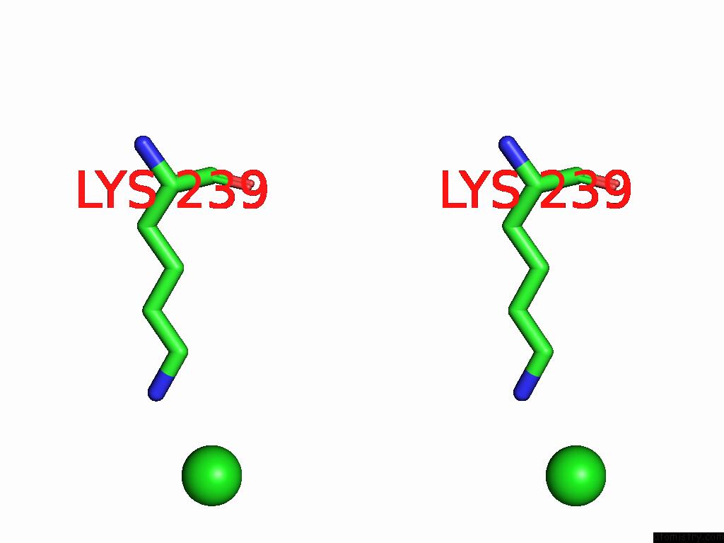
Stereo pair view
Mono view
Stereo pair view
A full contact list of Chlorine with other atoms in the Cl binding
site number 1 of Crystal Structure of the Coxiella Burnetii E110Q Mutant 2- Methylisocitrate Lyase within 5.0Å range:
|
Chlorine binding site 2 out of 2 in 9hgo
Go back to
Chlorine binding site 2 out
of 2 in the Crystal Structure of the Coxiella Burnetii E110Q Mutant 2- Methylisocitrate Lyase
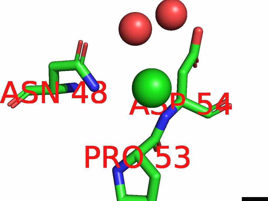
Mono view
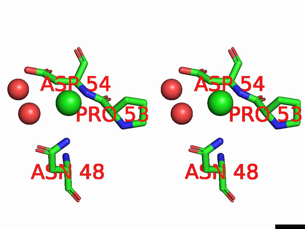
Stereo pair view
Mono view
Stereo pair view
A full contact list of Chlorine with other atoms in the Cl binding
site number 2 of Crystal Structure of the Coxiella Burnetii E110Q Mutant 2- Methylisocitrate Lyase within 5.0Å range:
|
Reference:
W.S.Stuart,
C.H.Jenkins,
P.M.Ireland,
M.N.Isupov,
I.H.Norville,
N.J.Harmer.
Structure and Catalytic Mechanism of Methylisocitrate Lyase, A Potential Drug Target Against Coxiella Burnetii. J.Biol.Chem. V. 301 08517 2025.
ISSN: ESSN 1083-351X
PubMed: 40250561
DOI: 10.1016/J.JBC.2025.108517
Page generated: Sat Aug 23 00:42:01 2025
ISSN: ESSN 1083-351X
PubMed: 40250561
DOI: 10.1016/J.JBC.2025.108517
Last articles
Mn in 9LJUMn in 9LJW
Mn in 9LJS
Mn in 9LJR
Mn in 9LJT
Mn in 9LJV
Mg in 9UA2
Mg in 9R96
Mg in 9VM1
Mg in 9P01