Chlorine »
PDB 9jl7-9lve »
9kk4 »
Chlorine in PDB 9kk4: Crystal Structure of Bovine Pancreatic Trypsin in Complex with Pyridoxine
Enzymatic activity of Crystal Structure of Bovine Pancreatic Trypsin in Complex with Pyridoxine
All present enzymatic activity of Crystal Structure of Bovine Pancreatic Trypsin in Complex with Pyridoxine:
3.4.21.4;
3.4.21.4;
Protein crystallography data
The structure of Crystal Structure of Bovine Pancreatic Trypsin in Complex with Pyridoxine, PDB code: 9kk4
was solved by
Z.Akbar,
M.S.Ahmad,
with X-Ray Crystallography technique. A brief refinement statistics is given in the table below:
| Resolution Low / High (Å) | 19.74 / 2.35 |
| Space group | P 21 21 21 |
| Cell size a, b, c (Å), α, β, γ (°) | 54.045, 57.75, 65.676, 90, 90, 90 |
| R / Rfree (%) | 19.9 / 26 |
Other elements in 9kk4:
The structure of Crystal Structure of Bovine Pancreatic Trypsin in Complex with Pyridoxine also contains other interesting chemical elements:
| Calcium | (Ca) | 1 atom |
Chlorine Binding Sites:
The binding sites of Chlorine atom in the Crystal Structure of Bovine Pancreatic Trypsin in Complex with Pyridoxine
(pdb code 9kk4). This binding sites where shown within
5.0 Angstroms radius around Chlorine atom.
In total 2 binding sites of Chlorine where determined in the Crystal Structure of Bovine Pancreatic Trypsin in Complex with Pyridoxine, PDB code: 9kk4:
Jump to Chlorine binding site number: 1; 2;
In total 2 binding sites of Chlorine where determined in the Crystal Structure of Bovine Pancreatic Trypsin in Complex with Pyridoxine, PDB code: 9kk4:
Jump to Chlorine binding site number: 1; 2;
Chlorine binding site 1 out of 2 in 9kk4
Go back to
Chlorine binding site 1 out
of 2 in the Crystal Structure of Bovine Pancreatic Trypsin in Complex with Pyridoxine
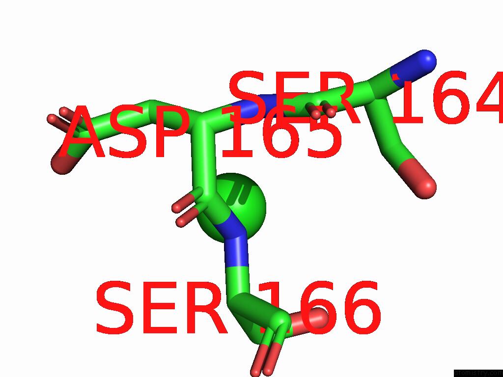
Mono view
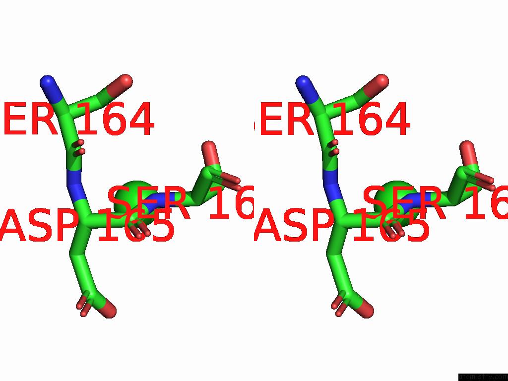
Stereo pair view
Mono view
Stereo pair view
A full contact list of Chlorine with other atoms in the Cl binding
site number 1 of Crystal Structure of Bovine Pancreatic Trypsin in Complex with Pyridoxine within 5.0Å range:
|
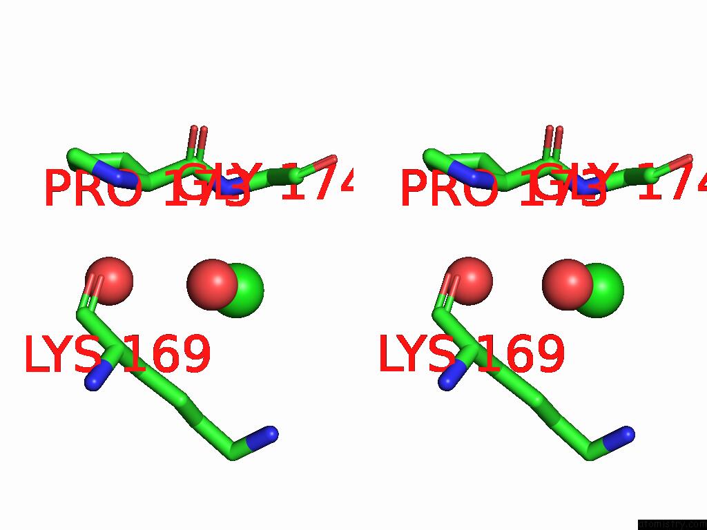
Chlorine binding site 2 out of 2 in 9kk4
Go back to
Chlorine binding site 2 out
of 2 in the Crystal Structure of Bovine Pancreatic Trypsin in Complex with Pyridoxine
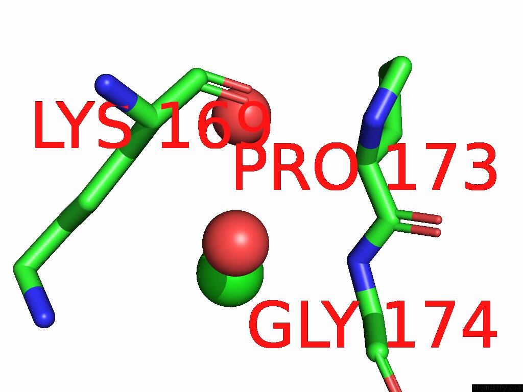
Mono view
Stereo pair view
Mono view
Stereo pair view
A full contact list of Chlorine with other atoms in the Cl binding
site number 2 of Crystal Structure of Bovine Pancreatic Trypsin in Complex with Pyridoxine within 5.0Å range:
|
Reference:
Z.Akbar,
N.Shah,
S.Mirza,
S.Rasheed,
M.S.Ahmad.
The in Vitro and Crystallographic Studies Reveal the Inhibitory Potential of Vitamin B 6 Analogues Against A Serine Protease Trypsin. Int.J.Biol.Macromol. V. 308 42433 2025.
ISSN: ISSN 0141-8130
PubMed: 40132700
DOI: 10.1016/J.IJBIOMAC.2025.142433
Page generated: Sun Jul 13 17:09:24 2025
ISSN: ISSN 0141-8130
PubMed: 40132700
DOI: 10.1016/J.IJBIOMAC.2025.142433
Last articles
Fe in 2YXOFe in 2YRS
Fe in 2YXC
Fe in 2YNM
Fe in 2YVJ
Fe in 2YP1
Fe in 2YU2
Fe in 2YU1
Fe in 2YQB
Fe in 2YOO