Chlorine »
PDB 1ahz-1bxz »
1bch »
Chlorine in PDB 1bch: Mannose-Binding Protein-A Mutant (Qpdwgh) Complexed with N- Acetyl-D-Galactosamine
Protein crystallography data
The structure of Mannose-Binding Protein-A Mutant (Qpdwgh) Complexed with N- Acetyl-D-Galactosamine, PDB code: 1bch
was solved by
A.R.Kolatkar,
W.I.Weis,
with X-Ray Crystallography technique. A brief refinement statistics is given in the table below:
| Resolution Low / High (Å) | 30.00 / 2.00 |
| Space group | C 1 2 1 |
| Cell size a, b, c (Å), α, β, γ (°) | 80.490, 85.010, 98.710, 90.00, 104.82, 90.00 |
| R / Rfree (%) | 21.9 / 25.2 |
Other elements in 1bch:
The structure of Mannose-Binding Protein-A Mutant (Qpdwgh) Complexed with N- Acetyl-D-Galactosamine also contains other interesting chemical elements:
| Calcium | (Ca) | 9 atoms |
| Sodium | (Na) | 1 atom |
Chlorine Binding Sites:
The binding sites of Chlorine atom in the Mannose-Binding Protein-A Mutant (Qpdwgh) Complexed with N- Acetyl-D-Galactosamine
(pdb code 1bch). This binding sites where shown within
5.0 Angstroms radius around Chlorine atom.
In total 3 binding sites of Chlorine where determined in the Mannose-Binding Protein-A Mutant (Qpdwgh) Complexed with N- Acetyl-D-Galactosamine, PDB code: 1bch:
Jump to Chlorine binding site number: 1; 2; 3;
In total 3 binding sites of Chlorine where determined in the Mannose-Binding Protein-A Mutant (Qpdwgh) Complexed with N- Acetyl-D-Galactosamine, PDB code: 1bch:
Jump to Chlorine binding site number: 1; 2; 3;
Chlorine binding site 1 out of 3 in 1bch
Go back to
Chlorine binding site 1 out
of 3 in the Mannose-Binding Protein-A Mutant (Qpdwgh) Complexed with N- Acetyl-D-Galactosamine
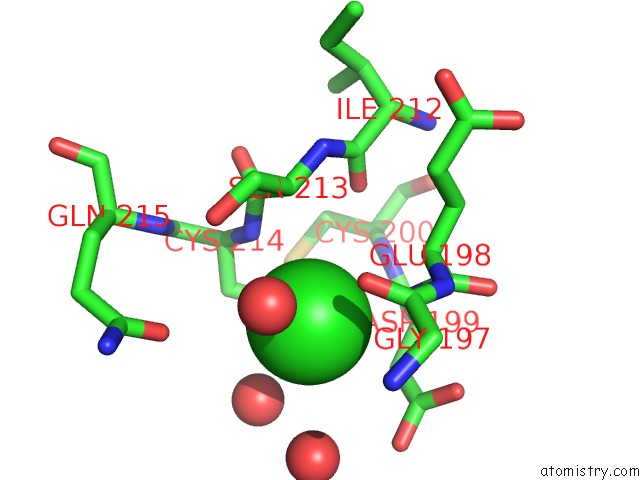
Mono view
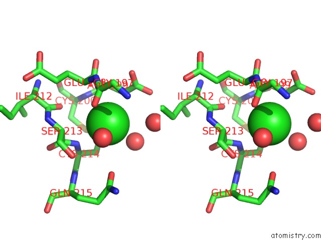
Stereo pair view
Mono view
Stereo pair view
A full contact list of Chlorine with other atoms in the Cl binding
site number 1 of Mannose-Binding Protein-A Mutant (Qpdwgh) Complexed with N- Acetyl-D-Galactosamine within 5.0Å range:
|
Chlorine binding site 2 out of 3 in 1bch
Go back to
Chlorine binding site 2 out
of 3 in the Mannose-Binding Protein-A Mutant (Qpdwgh) Complexed with N- Acetyl-D-Galactosamine
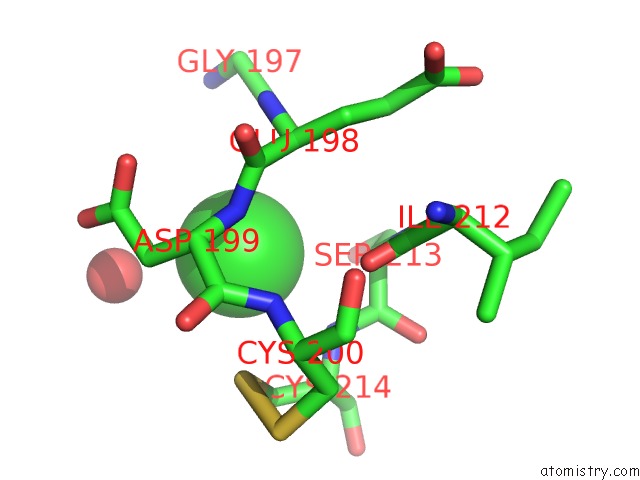
Mono view

Stereo pair view
Mono view
Stereo pair view
A full contact list of Chlorine with other atoms in the Cl binding
site number 2 of Mannose-Binding Protein-A Mutant (Qpdwgh) Complexed with N- Acetyl-D-Galactosamine within 5.0Å range:
|
Chlorine binding site 3 out of 3 in 1bch
Go back to
Chlorine binding site 3 out
of 3 in the Mannose-Binding Protein-A Mutant (Qpdwgh) Complexed with N- Acetyl-D-Galactosamine
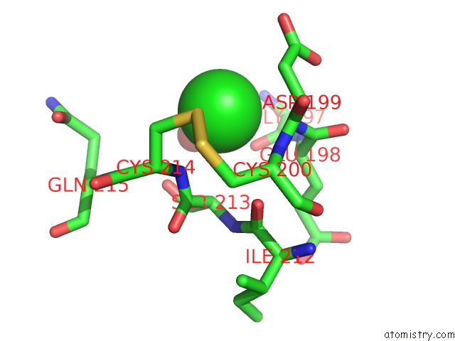
Mono view

Stereo pair view
Mono view
Stereo pair view
A full contact list of Chlorine with other atoms in the Cl binding
site number 3 of Mannose-Binding Protein-A Mutant (Qpdwgh) Complexed with N- Acetyl-D-Galactosamine within 5.0Å range:
|
Reference:
A.R.Kolatkar,
A.K.Leung,
R.Isecke,
R.Brossmer,
K.Drickamer,
W.I.Weis.
Mechanism of N-Acetylgalactosamine Binding to A C-Type Animal Lectin Carbohydrate-Recognition Domain. J.Biol.Chem. V. 273 19502 1998.
ISSN: ISSN 0021-9258
PubMed: 9677372
DOI: 10.1074/JBC.273.31.19502
Page generated: Thu Jul 10 16:20:12 2025
ISSN: ISSN 0021-9258
PubMed: 9677372
DOI: 10.1074/JBC.273.31.19502
Last articles
Cl in 5TR9Cl in 5TQU
Cl in 5TQJ
Cl in 5TPI
Cl in 5TQI
Cl in 5TPU
Cl in 5TPH
Cl in 5TPX
Cl in 5TPG
Cl in 5TOW